Abstract
Figures & Tables
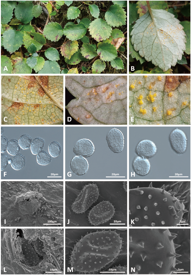
Fig. 1. occurring on . A, symptoms appeared on the upper surface of affected leaves. B, uredinia formed on the lower surface of an affected leaf. C-E, close-up view of erumpent uredinia. F-H, urediniospores observed under a DIC microscope: Urediniospores focused on outline (F and G), urediniospores focused on ornamentation (H). I-N, uredinium (I and L), urediniospores (J and M), and wall ornamentation of urediniospores (K and N) observed under a scanning electron microscope (SEM).