Intracellular Lipid Accumulation Inhibition, Anticancer Activity, and Single Oral Dose Toxicity of Ethanolic Wolfiporia cocos Extracts

 박나혜    이화용    최종운    박승춘*   

1Laboratory of Veterinary Pharmacokinetics & Pharmacodynamics, College of Veterinary Medicine, Kyungpook National University, Daegu 41566, Korea
2Department of Biology, Kunsan National University, Gunsan 54150, Korea
3Division of Food Technology, Biotechnology and Agrochemistry, Chonnam National University, Gwangju 61186, Korea

Abstract

In the present study, we compared the effects of 50% ethanolic extracts of Chinese and Korean Wolfiporia cocos (CPE and KPE) on in vitro lipid accumulation in 3T3-L1 cells and their anticancer activities in Sarcoma 180 cells. We further compared the anticancer activities and the 50% inhibitory concentrations (IC50) of CPE with KPE with cultivated for one and two years in a landfill and a facility (LPE and FPE), respectively. In addition, the single oral dose toxicities of CPE and KPE were evaluated in mice. Lipid accumulation was inhibited after 48 hours, in CPE and KPE treated 3T3-L1 cells; however, no significant difference was observed between CPE and KPE in their lipid accumulation inhibitory activities. The anticancer activity of KPE was higher than that of CPE at 300 μg/mL (p<0.05), revealing the possibility of an auxiliary biological means for origin identification. The anticancer activities of LPE and FPE were significantly stronger than that of CPE (p<0.05) but there was no difference between extracts from one- and two-year-old W. cocos, irrespective of the cultivation method. In single oral dose toxicity tests, CPE and KPE did not induce mortality during the 14-day observation. Thus, the 50% of lethal dose (LD50) of CPE and KPE were estimated to be higher than 2,000 mg/kg. Taken together, our results indicate that the anticancer assay could be an auxiliary means of identifying the origin of W. cocos. In addition, artificial cultivation could be an alternative way to reduce the import of W. cocos. Lastly, 50% ethanolic W. cocos extracts could be potential candidates for obesity and cancer managements.

Figures & Tables

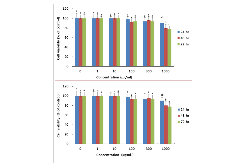
Fig. 1. Cell viability of 3T3-L1 cells 0.5 mM isobutylmethylxanthine (IBMX), 1 μM dexamethasone, 1 μg/mL insulin treated by Korean ethanolic extract (KPE) at the indicated concentration (0, 1, 10, 100, 300 and 1000 μg/mL) for 24, 48 and 72 hr. Growth rate was assessed by MTT assay. Data are expressed as percent growth rate of cells cultured in the presence of extract, compared with untreated control cells, taken 100%. All values are mean ± SD. Letters with different superscripts are significantly different by ANOVA with Duncan’s multiple range test atp < 0.05 at each time point.