Jin Sung Lee1†, Minkyeong Kim2†, Jae Young Park3, and Changmu Kim2*
1Yurim Mushroom Research Center, Asan 31582, Korea
2Species Diversity Research Division, National Institute of Biological Resources, Incheon 22689, Korea
3Q-myco, Seoul 08793, Korea
†These authors have contributed equally to this work.
*Correspondence to snubull@korea.kr
Korean Journal of Mycology (Kor J Mycol) 2025 December, Volume 53, Issue 4, pages 237-243.
https://doi.org/10.4489/kjm.2025.53.4.1
Received on October 17, 2025, Revised on November 10, 2025, Accepted on November 10, 2025, Published on December 31, 2025.
Copyright © The Korean Society of Mycology.
This is an Open Access article which is freely available under the Creative Commons Attribution-Non-Commercial 4.0 International License (CC BY-NC) (https://creativecommons.org/licenses/by-nc/4.0/).
Surveys of indigenous fungi were conducted in 2023 and 2024 around Magoksa Temple, located on the Taehwasan Mountain in Gongju-si. The species of all collected specimens were determined based on their morphological characteristics and rDNA sequences. Among these, Amanita yanshanensis, Cyclocybe erebioides, and Mallocybe aurantiodisca were newly identified as macromycota in Korea.
Macrofungal flora, Magoksa, Unrecorded species
The Magoksa Temple is located on the eastern hillside of the Taehwasan Mountain in Sagok-myeon, Gongju-si, Chungcheongnam-do, South Korea. It is registered as a World Heritage site due to being part of the Sansa Buddhist Mountain Monasteries in Korea [1]. The Magoksa Temple has a long history, and the surrounding forest system is well preserved. A variety of plants, including broad-leaved and coniferous trees, grew naturally in this area, resulting in the presence of a diverse range of higher fungi. Recently, the identification of macrofungi has been conducted using DNA sequences, and the number of newly identified and classified macrofungi has increased [2]. Following this trend, many new fungal species have been identified in Korea. In this study, both morphological observations and DNA sequence analysis of all the collected specimens were conducted, which led to the identification of new species of macrofungi that were previously categorized as the same species owing to morphological similarities. To distinguish between these species, detailed morphological characteristics were investigated. Surveys of higher fungi conducted in 2023 and 2024 revealed three species of agaric fungi that had not been previously recorded in Korea. This study presents three species that were newly recorded in Korea and provided comprehensive morphological descriptions of each species.
All specimens were primarily identified based on their macroscopic and microscopic features, with reference to previously published descriptions [3–7]. Measurements and images of the microscopic features were obtained using a Nikon Eclipse 80i microscope (Nikon, Tokyo, Japan). The sizes and shapes of more than 20 randomly selected basidiospores, basidia, and other microscopic features of each specimen were measured. For molecular identification, total DNA was extracted from specimen tissues collected on the same day using an AccuPrep Genomic DNA Extraction Kit (Bioneer, Daejeon, Korea). The internal transcribed spacer regions (ITS) were amplified using the primer set ITS1/ITS4 [8]. DNA sequencing was performed at the Macrogen DNA Sequencing Facility (Seoul, Korea) using the primers used for the PCR. Nucleotide sequences were edited using MEGA 11 software [9] and registered in GenBank. Species-level identification was conducted using reference sequences from GenBank obtained using BLASTn. Neighborjoining phylogenetic analysis was performed using MEGA 11 software with Jukes-Cantor correction. The robustness of the inferred neighbor-joining topology was tested using 1,000 bootstrap replicates. Newly identified fungal species were enumerated according to the current taxonomy by combining morphological and phylogenetic analysis results. Taxonomic classification and associated nomenclature of the species were assigned using the MycoBank database (http://www.mycobank.org/).
Three agaric species, Amanita yanshanensis Hao Zhou bis & C.L. Hou 2023, Cyclocybe erebioides Angelini & Vizzini 2014, and Mallocybe aurantiodisca Y.G. Fan, Y.P. Ge, J.H. Hu & W.J. Yu 2023 were observed in Korea for the first time. Here, we present photographs of fruiting bodies, drawings of microscopic features (Fig.1) and descriptions and discussion of these species. The sequences of these three species have been deposited in GenBank (accession numbers PP768066, PP768067, and PP768069). These sequences were compared with GenBank reference sequences (Table 1) and identified by ITS sequence analysis. The specimen NIBRFG0000515355 formed a monophyletic clade with the reference sequences of Amanita yanshanensis (bootstrap value = 100%; sequence similarity = 99.7–99.9%). NIBRFG0000515352 formed a monophyletic clade with reference sequences of Cyclocybe erebioides (bootstrap value = 100%, sequence similarity = 99.0–99.5%). The final specimen NIBRFG000515354 formed a monophyletic clade with the reference sequence, Mallocybe aurantiodisca (bootstrap value = 100%; sequence similarity = 99.2–99.7%) (Fig. 2).
Table 1. Closest GenBank matches of 3 undescribed species in this study
테일블
ITS: internal transcribed spacer regions.
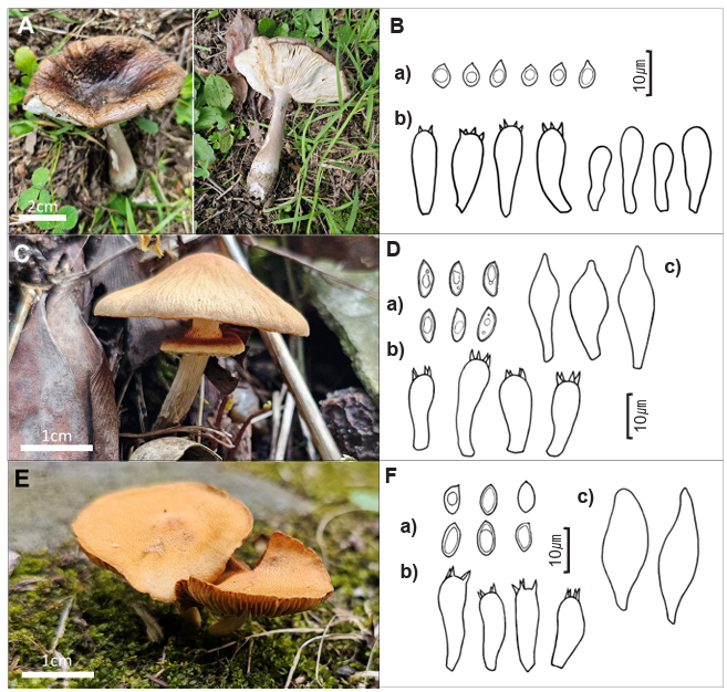
Fig. 1. Fruiting bodies and microscopic features of Amanita yanshanensis (A, B), Cyclocybe erebioides (C, D) and Mallocybe aurantiodisca (E, F). a, basidiospores; b, basidia & basidiol; c, Pleurocystidia. The scale bar is 10 μm in the microscopic images.
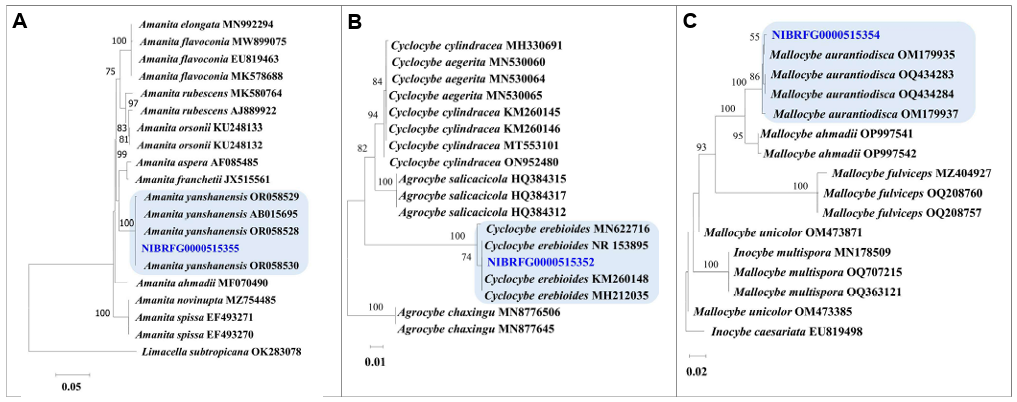
Fig. 2. Neighbor-joining tree of three unrecorded species constructed using the sequences of internal transcribed spacer regions. Bootstrap scores of >50 are presented at the nodes. The scale bar indicates the number of nucleotide substitutions per site. Amanita yanshanensis (A), Cyclocybe erebioides (B), and Mallocybe aurantiodisca (C).
Amanita yanshanensis Hao Zhou bis & C.L. Hou, in Zhou, Guo, Zhuo, Yan, Sui, Gao & Hou, IMA Fungus 14(no. 1226794): 20 (2023)
Korean name: Hoe-Jeok-Saek-Gwang-Dae-Beo-Seot; nom. nov. (회적색광대버섯) The Korean name refers to the color of the basidiomata
Pileus, 7 cm plano-convex to applanate, with an umbo at the center; the surface was dark grayish red, large, densely arranged over the disk; margin slightly striated; trama white. Lamellae, were free and crowded. Stipe, 7 cm long; 1 cm wide at apex; surface was gray-white with silk luster. Apical, subapical to fugacious; thin annulus; with cream color and silk luster on the upper surface; and matte cream-colored lower surface with floccose to farinose warts. Spore prints were not observed. Odor, indistinct.
Basidia 25–35 × 9–12 μm, clavate, 4-sterigmata, partially 2-sterigmata, without a basal clamp.
Basidiospores, 10–13 × 4.5–5 μm, subfusiform, and smooth. Pleurocystidia were 25–50 × 8–13 μm, fusiform, and thin-walled. Cheilocystidia, 31–45 × 9–15 μm, clavate to subfusiform, and thin-walled.
Specimen examined: Korea. Gongju-si, Magoksa Temple, 26 Aug 2023, NIBRFG0000515355 (JS20230826-24, GenBank accession No. PP768066)
Habitat: The soil found in deciduous forests.
Remarks: A. yanshanensis is well-circumscribed by its gray-white to dark grayish red pileus, which were densely arranged with pyramidal, subverrucose to subconical, floccose volval remnants on the stipe base arranged irregularly or in incomplete belts or rings. A. yanshanensis is very similar to A. flavoconia, sharing the common characteristic of producing spheroid to broadly ellipsoid, inamyloid spores. However, they show differences in the coloration of the pileus; A. yanshanensis exhibits a dark reddish-gray or grayish-brown hue, whereas A. flavoconia typically presents a yellowish or orange color [10].
Cyclocybe Velen., Novitates Mycologicae Novissimae: 122 (1940)
Korean name Min-Gat-Beo-Seot-Sok, nom. nov. (민갓버섯속)
The Korean name refers to the round smooth shape of the cap. Cyclocybe erebioides Angelini & Vizzini, in Vizzini, Angelini, and Ercole, Boll. Assoc. Micol. Ecol. Romana 92(2): 21–38 (2014)
Korean name: Ams-Saek- Min-Gat-Beo-Seot I, nom. nov. (암색민갓버섯)
The Korean name refers to the color of the basidiomata
Pileus, 22–37 mm in diameter; broadly convex when young and plane when mature; not clearly depressed or umbo, hygrophanous, viscid, glabrous, yellowish-brown to brown, and generally darker in center and fading toward the margin; and the margin were distinctly striated. Context was thin (up to 1 mm) and concolorous with a pileus surface. Lamellae were adnate or subdecurrent; thick; dense, with a series of 1–3 lamellulae; initially whitish and then light brown; and edges were distinctly white. Stipe, 40 mm × 3 mm; central; cylindrical; hollow; dry; fibrous; with an annular zone; initially whitish; darken with age; or brown at the touch. Annulus, single; white; membranous; persistent; and upturned. Odor, not distinctive
Basidia, 17–35 × 6–10 μm; clavate, thin-walled; and 4-sterigmata. Basidiospores, 9–11 × 4–5 μm; subfusiform to ellipsoid; slightly thick-walled; and smooth. Pleurocistidia, 35–70 × 7–12 μm; subclavate or subfusiform; ventricose; and thin-walled.
Specimen examined: Korea. Gongju-si, Magoksa Temple, 26 Aug 2023, NIBBRFG0000515352 (JS20230826-22, GenBank accession No. PP768067)
Habitat: In the soil of deciduous forests.
Remarks: Cyclocybe erebioides is characterized by a wide and complex membranous annulus with clamp-connections. C. erebioides is similar to C. erebia, sharing the characteristics of having smooth basidiospores and a well-developed membranous annulus. However, these 2 species differ slightly in the size and morphology of their spores [11].
Mallocybe aurantiodisca Y.G. Fan, Y.P. Ge, J.H. Hu & W.J. Yu, in Hu, Yu, Deng, Fan, Bau, Tang, Lin & Deng, Mycol. Progr. 22(2, no. 15): 13 (2023)
Korean name. Ju-Hong-Yang-Teol-Gat-Beo-Seot, nom. nov. (주홍양털갓버섯)
The Korean name refers to the color of the basidiomata
Pileus small-sized (13–25 mm in diameter); convex to broadly convex; mentose to woolly tomentose with scurfy appressed squamules; and brownish with orange tinge. Lamellae, 1–3 mm wide; adnate; narrow; and ochraceous brown in color. Stipe, 30 × 3 mm, cylindrical or slightly tapering toward base, pale yellow, with appressed velar remnants, and white. Context creamy white or pale yellowish white in pileus, 1–2 mm thick, and concolorous in stipe.
Basidia, 31–45 × 8–10 μm; clavate; and 4-sterigmata. Basidiospores, 7.5–9.8 × 3.5–4.6 μm; subfusiform; and brownish yellow. Pleurocystidia, 47–93 × 10–17 μm; clavate or ventricose; and thinwalled.
Specimen examined: Korea. Gongju-si, Magoksa Temple, 26 Aug 2023, NIBRFFG0000515354 (JS20230826-23, GenBank accession No. PP768069)
Habitat: Soil covered with moss.
Remarks: Mallocybe aurantiodisca is characterized by its reddish orange-tinged basidiomata; tomentosesquamulose pileus; elongate-ellipsoid basidiospores; utriform; and ventricose to cylindrical cheilocystidia that are usually flexuous. M. aurantiodisca and M. himalayana share morphological similarities, including an ectomycorrhizal lifestyle and the production of bean-shaped basidiospores. However, M. aurantiodisca may occasionally display a yellowish tint at the stipe base, a feature consistently absents in M. himalayana, which has a more uniformly fibrous stipe [12].
Most previous studies on macrofungi focused on national parks, therefore this survey is significant because we concentrated on mountainous temple areas in the central region of South Korea, which are relatively understudied. Despite the short survey period, three previously unrecorded species were identified in Korea. Advances in molecular biology have contributed to the identification of these fungal species, specifically genealogical concordance phylogenetic species recognition, which has helped improve understanding of the relationships among macrofungi [13].
The three previously unrecorded species identified in this survey belong to the genera Amanita Pers., Cyclocybe Velen, and Mallocybe (Kuyper) Vizzini. The genus Amanita is characterized by a cap and stipe with both universal and partial veils, often forming a volva and ring. They include both highly toxic and edible species, making accurate identification essential. Most species are ectomycorrhizal and commonly found in forested areas [10,14]. The genus Amanita has been relatively well documented in Korea, with approximately 80 species reported to date [15]. Amanita yanshanensis has previously only been reported in southern and southwestern China [10]. Its newly identified presence in Korea suggests the possibility of a northward shift in its natural habitat owing to climate change (global warming). With more detailed observations, its designation as an indicator species of climate change may be worth considering. Cyclocybe is primarily a saprotrophic fungal genus that decomposes wood. Mushrooms are generally small to medium in size, and some species are edible or even cultivated. In the past, certain species were classified under the genus Agrocybe, but molecular phylogenetic studies have led to their reclassification under Cyclocybe [11,16]. In Korea, 2 species of Cyclocybe have been confirmed: Cyclocybe erebia (Fr.) (Vizzini & Matheny) and C. cylindracea (DC.) (Vizzini & Matheny). C. erebioides was the third identified species of the genus Cyclocybe in Korea [14]. Mallocybe is a genus of small mushrooms, which is primarily found in subtropical and temperate regions. Previously, species in this genus were included in the genus Inocybe, but molecular phylogenetic studies have resulted in their separation into distinct genera. They are mainly mycorrhizal fungi that grow in the soil. Their caps are small, bell-shaped, and expand gradually as they mature. The colors are typically brown, yellowish-brown, or gray, and the surface may have fibrous patterns or scales [17]. Mallocybe aurantiodisca was the second species of the genus Mallocybe to be reported in Korea after M. malenconii [14].
Since fungi have a limited period during which fruiting bodies form, collecting and observing them can be challenging. To address this issue, long-term collection surveys in specific regions are required and more species diversity is expected to be uncovered through selective and intensive research.
This study was supported by a grant from the National Institute of Biological Resources (NIBR) of the Republic of Korea (NIBR202502103).
1. Gongju City. Magoksa Temple. Gongju: Gongju City; 2020.
2. Laurence MH, Summerell BA, Burgess LW, Liew EC. Genealogical concordance phylogenetic species recognition in the Fusarium oxysporum species complex. Fungal Biol 2014;118:374-84. https://doi.org/10.1016/j.funbio.2014.02.002
[DOI]
3. Breitenbach J, Kränzlin F. The fungi of Switzerland. Vols. 1-5. Lucerne: Verlag Mykologia; 1984-2000.
4. Gilbertson RL, Ryvarden L. North American polypores. Vol. 1. Abortiporus-Lindtneria. Oslo: Fungiflora; 1986.
5. Hongo T, Izawa M. Yama-Kei field book. No. 10. Fungi. Tokyo: Yama-Kei Publishers; 1994.
6. Imazeki R, Otani Y, Hongo T, Izawa M, Mizuno N. Fungi of Japan. Tokyo: Yama-Kei Publishers; 1988.
7. Kränzlin F. Fungi of Switzerland. Vol. 6. Russulaceae: Lactarius, Russula. Lucerne: Verlag Mykologia; 2005.
8. White TJ, Bruns TD, Lee SB, Taylor JW. Amplification and direct sequencing of fungal ribosomal RNA genes for phylogenetics. In: Innis MA, Gelfand DH, Sninsky JJ, editors. PCR protocols: A guide to methods and applications. New York: Academic Press; 1990. p. 315-22.
[DOI]
9. Tamura K, Peterson D, Peterson N, Stecher G, Nei M, Kumar S. MEGA11: Molecular evolutionary genetics analysis using maximum likelihood, evolutionary distance, and maximum parsimony methods. Mol Biol Evol 2011;28:2731-9. https://doi.org/10.1093/molbev/msr121
[DOI]
10. Zhou H, Guo M, Zhuo L, Yan H, Sui X, Gao Y, Hou C. Diversity and taxonomy of the genus Amanita (Amanitaceae, Agaricales) in the Yanshan Mountains, Northern China. Front Plant Sci 2023;14:1226794. https://doi.org/10.3389/fpls.2023.1226794
[DOI]
11. Vizzini A, Angelini C, Ercole E. Le sezioni Velatae e Aporus di Agrocybe sottogenere Aporus: Rivalutazione del genere Cyclocybe Velen. ed una nuova specie. Rivista Micol Romana 2014;92:21-38.
12. Hu JH, Yu WJ, Deng LS, Fan YG, Bau T, Tang LP, Lin WF, Deng CY. The detection of major clades and new species of Mallocybe (Inocybaceae, Agaricales) from China with elongate cheilocystidia. Mycol Prog 2023;22:15. https://doi.org/10.1007/s11557-022-01854-5
[DOI]
13. Taylor JW, Jacobson DJ, Kroken S, Kasuga T, Geiser DM, Hibbett DS, Fisher MC. Phylogenetic species recognition and species concepts in fungi. Fungal Genet Biol 2000;31:21-32. https://doi.org/10.1006/fgbi.2000.1228
[DOI]
14. Singer R. The Agaricales in modern taxonomy. 4th ed. Koenigstein: Koeltz; 1986.
15. National Institute of Biological Resources (NIBR). National species list of Korea. Incheon: NIBR; 2024.
16. Zhu XY, Nyima T, Vizzini A, He MQ, Phurbu D, Zhao RL. Two new species of Cyclocybe (Agaricales, Tubariaceae) from China. Phytotaxa 2023;620:33-46. https://doi.org/10.11646/phytotaxa.620.1.3
[DOI]
17. Matheny PB, Kudzma LV. New species of Inocybe (Inocybaceae) from eastern North America. J Torrey Bot Soc 2019;146:213-35. https://doi.org/10.3159/TORREY-D-18-00060.1
[DOI]