
Thuong T. T. Nguyen, and Hyang Burm Lee*
Environmental Microbiology Laboratory, Department of Agricultural Biological Chemistry, College of Agriculture & Life Sciences, Chonnam National University, Gwangju 61186, Korea
*Correspondence to hblee@jnu.ac.kr
Korean Journal of Mycology (Kor J Mycol) 2026 June, Volume 54, Issue 2, pages 83-89.
https://doi.org/10.4489/kjm.2026.54.2.1
Received on April 16, 2026, Revised on April 27, 2026, Accepted on April 29, 2026, Published on June 30, 2026.
Copyright © The Korean Society of Mycology.
This is an Open Access article which is freely available under the Creative Commons Attribution-Non-Commercial 4.0 International License (CC BY-NC) (https://creativecommons.org/licenses/by-nc/4.0/).
Eurotiales, Penicillago, Phylogeny, Taxonomy
Eurotiales is a relatively large order of ascomycete fungi comprising species that have been widely used in food fermentation and biotechnology for the production of enzymes, organic acids, and pharmaceuticals. However, some species in this order are also associated with food spoilage, mycotoxin production, indoor contamination, and opportunistic infections [1–4].
At present, Eurotiales comprises four families (Aspergillaceae, Penicillaginaceae, Thermoascaceae and Trichocomaceae), 26 genera (Acidotalaromyces, Ascospirella, Aspergillago, Aspergillus, Dendrosphaera, Evansstolkia, Hamigera, Leiothecium, Monascus, Paecilomyces, Penicillago, Penicilliopsis, Penicillium, Phialomyces, Pseudohamigera, Pseudopenicillium, Rasamsonia, Sagenomella, Sclerocleista, Talaromyces, Thermoascus, Thermomyces, Trichocoma, Warcupiella, Xerochrysium and Xeromyces), and 1,393 species [5].
The genus Penicillago (Penicillaginaceae, Eurotiales) was introduced to accommodate the taxonomically confusing species Penicillium nodositatum [6]. Subsequently, three additional species were added: P. kabunica, P. mirabilis, and P. moldavica [7]. Four species, P. kabunica, P. mirabilis, P. moldavica, and P. nodositata, are currently listed in Index Fungorum (http://www.indexfungorum.org/names/names.asp) [8]. However, to date, no species of this genus has been reported in Korea.
Morphologically, species of Penicillago are characterized by the production of Penicillium-like conidiophores, which contain ampulliform phialides with relatively long, narrow necks and green conidia. They are unable to grow on Czapek yeast autolysate agar (CYA) when incubated at 37°C and exhibit moderate growth on creatine sucrose agar (CREA) without acid production [6,7].
During our investigation of fungal diversity in soil in Korea, we identified a newly recorded species, Penicillago mirabilis, belonging to the order Eurotiales, based on morphological characteristics and multi-gene phylogenetic analyses. Descriptions, illustrations, and results of phylogenetic analyses of this species are provided.
Soil samples were collected from Ulleung Island, Korea (37°30′23.0″N 130°51′25.9″E). One gram of soil was mixed with 9 mL sterile distilled water. Serial dilutions of the mixtures were then prepared (from 10−1 to 10−5). For each dilution, a 100 µL aliquot was dispensed onto potato dextrose agar (PDA; Becton, Dickinson and Co., Sparks, MD, USA) supplemented with neomycin (50 mg/L). The plates were incubated at 25°C in the dark for 7 days. Individual fungal colonies were transferred to new PDA plates and incubated at 25°C for 3–7 days. Pure isolates were obtained and stored on PDA slants at 4°C and in 20% glycerol at −80°C at the Environmental Microbiology Laboratory Fungarium, Chonnam National University, Gwangju, Korea. The isolated strain was also deposited in the Collection of National Institute of Biological Resources (NIBR), Incheon, Korea.
Three-point inoculations were performed on CYA, malt extract agar (MEA), yeast extract sucrose agar (YES), oatmeal agar (OA), and CREA [9,10]. All media were incubated at the standard temperature of 25°C for 7 days. Additional plates were incubated at 30 and 37°C. Macroscopic and microscopic features were studied after 7 days. Slides were prepared using 60% lactic acid, and excess conidia were removed using 96% ethanol. An Olympus BX53 microscope with differential interference contrast optics (Olympus, Tokyo, Japan) was used to obtain digital images.
Fresh mycelium was scraped from the margins of colonies on PDA overlaid with cellophane after 7 days incubated at 25°C. Genomic DNA was extracted using a SolgTM Genomic DNA Prep Kit (Solgent Co. Ltd., Daejeon, Korea) according to the manufacturer’s instructions. The internal transcribed spacer (ITS) region of rRNA, β-tubulin (BenA), calmodulin (CaM), and RNA polymerase II second largest subunit (RPB2) were amplified using primers V9G/ITS4 [11,12], Bt2a/Bt2b [13], CF1L/CF4 [14], and RPB2-5F/RPB2-7cR [15], respectively. The PCR products were purified using an AccuPrep PCR Purification Kit (Bioneer, Daejeon, Korea). DNA sequencing was performed at Macrogen (Daejeon, South Korea) using the same primers used for PCR.
The generated sequences, together with reference sequences for closely related species based on the BLAST results, were aligned using MAFFT v. 7 (http://mafft.cbrc.jp/alignment/server/) [16] and then manually edited in MEGA 7 [17]. Phylogenetic analyses were based on maximum likelihood performed in IQ-TREE 2 and bootstrap analyses using 1,000 replicates [18]. The tree figures were created using FigTree v1.3.1. Bootstrap support values were higher than 70% in the branches. All sequences generated in this study were deposited in GenBank (https://www.ncbi.nlm.nih.gov) (Table 1).
Table 1. Strains and GenBank accession numbers used for phylogenetic analysis in this study
| Species | Strain no. | GenBank accession number | |||
|---|---|---|---|---|---|
| ITS | BenA | CaM | RPB2 | ||
| Penicillago kabunica | CBS 575.90T | MN431415 | MN969438 | MN969357 | MN969217 |
| Penicillago mirabilis | CBS 624.72T | MN431416 | MN969439 | MN969358 | MN969218 |
| Penicillago mirabilis | CNUFC U8-51 | PZ327461 | PZ338222 | PZ338223 | PZ338224 |
| Penicillago moldavica | CBS 574.90T | MN431417 | MN969440 | MN969359 | MN969219 |
| Penicillago nodositata | CBS 333.90T | KC790403 | KC790399 | MN969361 | MN969220 |
| Penicillago sp. | FMR 16442 | LT899788 | LT898313 | LT899772 | LT899806 |
| Penicillago sp. | FMR 15296 | LT899787 | LT898312 | LT899771 | LT899805 |
| Penicillago sp. | DTO 119-A2 | N/A | N/A | MT066181 | MT066178 |
| Penicillago sp. | DTO 216-G1 | N/A | MF974887 | MT066182 | MT066179 |
Bold letters indicate strains and accession numbers determined in this study. ITS: internal transcribed spacer; BenA: β-tubulin; CaM: calmodulin; RPB2: RNA polymerase II second largest subunit; CBS: Culture collection of the Westerdijk Fungal Biodiversity Institute, The Netherlands; CNUFC: Chonnam National University Fungal Collection, Gwangju, Korea; DTO: Internal Culture Collection of the CBS-Fungal Biodiversity Center; FMR: Facultat de Medicina i Ciències de la Salut, Reus, Spain; N/A: not available. ᵀex-type strain.
Phylogenetic analyses of Penicillago species were performed using a concatenated dataset of four loci (BenA, CaM, ITS, and RPB2) (Fig. 1). The multi-gene analysis included nine taxa. The concatenated alignment consisted of 2,880 characters (including alignment gaps): 481, 527, 895, and 977 characters were used in BenA, CaM, ITS, and RPB2, respectively. Multi-gene phylogenetic analysis revealed that our strain (CNUFC U8-51) clustered with an ex-type strain of Penicillago mirabilis (Fig. 1).
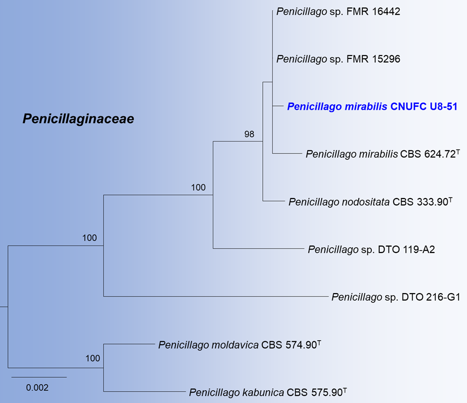
Fig. 1. Phylogram generated using maximum likelihood (ML) analysis based on combined β-tubulin (BenA), calmodulin (CaM), internal transcribed spacer (ITS), and RNA polymerase II second largest subunit (RPB2) sequence data for Penicillago species. Branches with >70% ML bootstrap support are indicated. Penicillago kabunica CBS 575.90 and P. moldavica CBS 574.90 were used as the outgroups. The newly generated sequences are indicated in bold blue. Ex-type strains are marked with a superscript T.
Penicillago mirabilis (Beliakova & Milko) Houbraken, Frisvad & Samson, Stud. Mycol. 95: 90 (2020) (Fig. 2).
Culture characteristics (7 days at 25°C): On CYA, colonies radially sulcate and protuberant at the center, mycelia white, texture velutinous, sporulation sparse, soluble pigments or exudates absent, reverse moderate yellowish pink. On MEA, colonies plane, texture floccose, mycelia white, sporulation moderate to dense, soluble pigments or exudates absent, reverse pale yellow. On YES, concentric rings toward the center, texture velutinous, mycelia white, sporulation very sparse or absent, soluble pigments or exudates absent, reverse yellowish brown and white toward the edges. On OA, colonies slightly raised at the center and cottony, mycelia white, sporulation sparse, soluble pigments or exudates absent, reverse white. On CREA, acid production absent.
Micromorphology: Conidiophores biverticillate. Stipes rough walled, 198–432 × 2.5–3.5 µm. Metulae three to five, divergent, 7.5–10.5 × 2.5–3.5(–4) µm. Phialides ampulliform, 6.5–10 × 2.5–3.5 µm. Conidia echinulate walled, subglobose, oval to broad fusiform, 2.6–3.0 × 2.5–2.8 µm. Ascomata not observed.
Colony diameters (mm), 7 days: CYA 25°C, 40–43; CYA 30°C, 24–26; CYA 37°C, no growth; MEA 25°C, 49–51; YES 25°C, 35–37; OA 25°C, 26–30; CREA 25°C, 26–31.
Material examined: Republic of Korea, Ulleung Island (37°30′23.0″N 130°51′25.9″E); from rhizosphere soil; May 1, 2019, culture CNUFC U8-51= NIBRFG0000509589.
Notes: Phylogenetic analysis of the combined BenA, CaM, ITS, and RPB2 dataset indicates that our strain CNUFC U8-51 is most closely related to P. mirabilis. Pairwise comparisons between CNUFC U8-51 and the reference strain of P. mirabilis showed high sequence similarity among all loci examined, with identities of 565/566 bp (99.82%) for ITS, 436/437 bp (99.77%) for BenA, 490/491 bp (99.79%) for CaM, and 767/768 bp (99.87%) for RPB2.
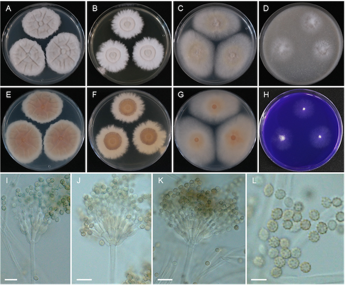
Fig. 2. Penicillago mirabilis (CNUFC U8-51). A, E: Colonies on Czapek yeast autolysate agar; B, F: Colonies on yeast extract sucrose agar; C, G: Colonies on malt extract agar; D: Colonies on oatmeal agar; H: Colonies on creatine sucrose agar. A–D, H: Obverse view; E–G: reverse view. I–K: Conidiophores; L: Conidia. Scale bars: I–K = 10 µm, L = 5 µm.
In the present study, we identified a rare species, Penicillago mirabilis, as a newly recorded species in Korea. This finding contributes to a broader understanding of the fungal diversity of Eurotiales in Korea.
Based on our multi-locus phylogenetic analysis, our strain CNUFC U8-51 is most closely related to P. mirabilis. The results of our molecular data analysis are consistent with the phylogeny presented by Houbraken et al. [7]. Morphologically, CNUFC U8-51 is consistent with the original description of P. mirabilis. However, our strain shows slower colony diameter growth on YES than that of P. mirabilis (35–37 mm vs. 51–59 mm) [7]. Based on the molecular and morphological evidence together, we identify strain CNUFC U8-51 as P. mirabilis. This isolate was obtained from a soil sample, thereby expanding the known ecological range of the species and contributing additional molecular data for the genus Penicillago.
The ITS region has been proposed as a universal DNA barcode marker for fungi [19]. However, ITS rDNA alone is not sufficient to distinguish all species in Eurotiales. Therefore, BenA and CaM have been proposed as alternative markers [7,9,20]. Even with additional gene regions, this study shows that more may be required, as illustrated by the taxonomy of P. mirabilis and P. nodositata.
The authors declare no conflicts of interest.
This study was partly supported by the project (2024-0153-01) of Chonnam National University and also by the Survey and Discovery of Indigenous Fungal Species of Korea Project, funded by the National Institute of Biological Resources (NIBR).
1. Houbraken J, de Vries RP, Samson RA. Modern taxonomy of biotechnologically important Aspergillus and Penicillium species. Adv Appl Microbiol 2014;86:199-249. https://doi.org/10.1016/b978-0-12-800262-9.00004-4
[DOI][PubMed]
2. Frisvad JC, Hubka V, Ezekiel CN, Hong SB, Nováková A, Chen AJ, Arzanlou M, Larsen TO, Sklenář F, Mahakarnchanakul W, et al. Taxonomy of Aspergillus section Flavi and their production of aflatoxins, ochratoxins and other mycotoxins. Stud Mycol 2019;93:1-63. https://doi.org/10.1016/j.simyco.2018.06.001
[DOI][PMC]
3. Pitt JI, Hocking AD. Fungi and food spoilage. 3rd ed. New York: Springer; 2009. p. 145-68. https://doi.org/10.1007/978-0-387-92207-2
[DOI]
4. van den Brule T, Punt M, Teertstra W, Houbraken J, Wösten H, Dijksterhuis J. The most heat-resistant conidia observed to date are formed by distinct strains of Paecilomyces variotii. Environ Microbiol 2020;22(3):986-99. https://doi.org/10.1111/1462-2920.14791
[DOI][PMC]
5. Visagie CM, Houbraken J, Overy DP, Sklenář F, Bensch K, Frisvad JC, Mack J, Perrone G, Samson RA, van Vuuren NI, et al. From chaos to tranquillity: A modern approach to the identification, nomenclature and phylogeny of Aspergillus, Penicillium and other Eurotiales, including an updated accepted species list. Stud Mycol 2025;112:117-260. https://doi.org/10.3114/sim.2025.112.04
[DOI][PMC]
6. Guevara-Suarez M, García D, Cano-Lira JF, Guarro J, Gené J. Species diversity in Penicillium and Talaromyces from herbivore dung, and the proposal of two new genera of penicillium-like fungi in Aspergillaceae. Fungal Syst Evol 2020;5:39-75. https://doi.org/10.3114/fuse.2020.05.03
[DOI][PMC]
7. Houbraken J, Kocsubé S, Visagie CM, Yilmaz N, Wang XC, Meijer M, Kraak B, Hubka V, Bensch K, Samson RA, et al. Classification of Aspergillus, Penicillium, Talaromyces and related genera (Eurotiales): An overview of families, genera, subgenera, sections, series and species. Stud Mycol 2020;95:5-169. https://doi.org/10.1016/j.simyco.2020.05.002
[DOI][PubMed][PMC]
8. Index Fungorum. Index Fungorum [Internet]. Kew: The Royal Botanic Gardens; 2025 [cited 2026 Mar 1]. Available from http://www.indexfungorum.org
9. Samson RA, Visagie CM, Houbraken J, Hong SB, Hubka V, Klaassen CHW, Perrone G, Seifert KA, Susca A, Tanney JB, et al. Phylogeny, identification and nomenclature of the genus Aspergillus. Stud Mycol 2014;78:141-73. https://doi.org/10.1016/j.simyco.2014.07.004
[DOI][PMC]
10. Visagie CM, Houbraken J, Frisvad JC, Hong SB, Klaassen CHW, Perrone G, Seifert KA, Varga J, Yaguchi T, Samson RA. Identification and nomenclature of the genus Penicillium. Stud Mycol 2014;78: 343-71. https://doi.org/10.1016/j.simyco.2014.09.001
[DOI][PubMed][PMC]
11. de Hoog GS, Gerrits van den Ende AH. Molecular diagnostics of clinical strains of filamentous Basidiomycetes. Mycoses 1998;41(5-6):183-9. https://doi.org/10.1111/j.1439-0507.1998.tb00321.x
[DOI]
12. White TJ, Bruns T, Lee S, Taylor J. Amplification and direct sequencing of fungal ribosomal RNA genes for phylogenetics. In: Innis MA, Gelfand DH, Sninsky JJ, White TJ, editors. PCR protocols: A guide to methods and applications. San Diego: Academic Press, 1990. p. 315-22. https://doi.org/10.1016/B978-0-12-372180-8.50042-1
[DOI][PubMed][PMC]
13. Glass NL, Donaldson GC. Development of primer sets designed for use with the PCR to amplify conserved genes from filamentous ascomycetes. Appl Environ Microbiol 1995;61(4):1323-30. https://doi.org/10.1128/aem.61.4.1323-1330.1995
[DOI][PubMed][PMC]
14. Peterson SW. Phylogenetic analysis of Aspergillus species using DNA sequences from four loci. Mycologia 2008;100(2):205-26. https://doi.org/10.3852/mycologia.100.2.205
[DOI][PubMed]
15. Liu YJ, Whelen S, Hall BD. Phylogenetic relationships among ascomycetes: Evidence from an RNA polymerase II subunit. Mol Biol Evol 1999;16(12):1799-808. https://doi.org/10.1093/oxfordjournals.molbev.a026092
[DOI]
16. Katoh K, Rozewicki J, Yamada KD. MAFFT online service: Multiple sequence alignment, interactive sequence choice and visualization. Brief Bioinform 2019;20(4):1160-6. https://doi.org/10.1093/bib/bbx108
[DOI][PubMed][PMC]
17. Kumar S, Stecher G, Tamura K. MEGA7: Molecular evolutionary genetics analysis version 7.0 for bigger datasets. Mol Biol Evol 2016;33:1870-4. https://doi.org/10.1093/molbev/msw054
[DOI][PMC]
18. Minh BQ, Schmidt HA, Chernomor O, Schrempf D, Woodhams MD, von Haeseler A, Lanfear R. IQ-TREE 2: New models and efficient methods for phylogenetic inference in the genomic era. Mol Biol Evol 2020;37(5):1530-4. https://doi.org/10.1093/molbev/msaa015
[DOI][PubMed][PMC]
19. Schoch CL, Seifert KA, Huhndorf S, Robert V, Spouge JL, Levesque CA, Chen W, Fungal Barcoding Consortium. Nuclear ribosomal internal transcribed spacer (ITS) region as a universal DNA barcode marker for Fungi. Proc Natl Acad Sci USA 2012;109(16):6241-6. https://doi.org/10.1073/pnas.1117018109
[DOI][PMC]
20. Yilmaz N, Visagie CM, Houbraken J, Frisvad JC, Samson RA. Polyphasic taxonomy of the genus Talaromyces. Stud Mycol 2014;78:175-341. https://doi.org/10.1016/j.simyco.2014.08.001
[DOI][PubMed][PMC]