
Jae Young Park1, Do Gyu Choi1, Sun Young Choi2, and Jin Sung Lee2,*
1Q-myco, Seoul 08793, Korea
2Yurim Mushroom Research Center, Asan 31582, Korea
*Correspondence to jsmush@naver.com
Korean Journal of Mycology (Kor J Mycol) 2026 June, Volume 54, Issue 2, pages 139-154.
https://doi.org/10.4489/kjm.2026.54.2.4
Received on April 13, 2026, Revised on May 22, 2026, Accepted on June 01, 2026, Published on June 30, 2026.
Copyright © The Korean Society of Mycology.
This is an Open Access article which is freely available under the Creative Commons Attribution-Non-Commercial 4.0 International License (CC BY-NC) (https://creativecommons.org/licenses/by-nc/4.0/).
Fungal diversity, Molecular phylogeny, New records, Songnisan National Park, Taxonomy
South Korea, which is characterized by its complex topography and distinct seasonal climatic conditions, has a highly favorable environment for fungal biodiversity. However, despite this ecological potential, mycological research in the region remains insufficient. Approximately 35,000 macrofungal species have been formally described worldwide; however, the number of macromycetes documented in Korea is estimated to be less than 2,200 species, representing less than 10% of the recorded global diversity [1,2]. This significant gap underscores the urgent need for comprehensive surveys to uncover Korea’s underexplored mycodiversity. Understanding fungal diversity is not only essential for mapping the complete ecological landscape but also for identifying potential novel species and new geographical records. In this context, national parks, such as Songnisan National Park, serve as critical repositories of biodiversity, offering well-preserved natural environments and diverse forest types that are ideal for the discovery of previously undocumented fungal taxa [3].
Songnisan National Park, a pivotal axis of the Baekdudaegan mountain range in central Korea, encompasses a total area of 274.5 km2 and is characterized by rugged granite topography and diverse forest ecosystems. The vegetation predominantly comprises mixed deciduous broad-leaved and coniferous forests, with Quercus and Pinus species being the dominant taxa. An average annual temperature of 11.9℃ and annual precipitation of 1,300 mm contribute to the maintenance of high humidity levels during the summer monsoon season, providing an optimal microclimate that enables diverse macromycetes to flourish [4]. This environmental complexity, coupled with well-preserved organic litter layers, establishes Songnisan as a critical repository for indigenous fungal biodiversity and a primary site for mycological research in the Korean Peninsula [3].
Previous mycological studies in Songnisan National Park date back to the 1990s [5,6]. The comprehensive resource surveys conducted by the Korea National Park Service (KNPS) have significantly contributed to the expansion of the local fungal checklist [4,7]. However, previous studies have primarily relied on morphological characteristics. In recent integrative taxonomic approaches, the diversity of Korean macromycetes is being re-evaluated, highlighting the necessity for updated surveys such as those conducted in 2024 [4].
Recent advancements in molecular systematics, particularly the utilization of the internal transcribed spacer (ITS) region as a standard DNA barcode, have significantly enhanced the precision of species identification. These modern molecular techniques complement traditional morphological analyses, thereby enabling a more robust and integrative approach to taxonomic studies.
A comprehensive mycological survey was conducted at Songnisan National Park from 2024 to 2025, leading to the identification of a total of 253 species belonging to 100 genera, 40 families, 15 orders, 5 classes, and 2 phyla. Among the collected specimens, six species—Aureoboletus miniatoaurantiacus, Boletus bainiugan, Lactarius mirus, Phaeotremella yunnanensis, Ramaria rubella, and Xerocomus galbanus—were confirmed to be newly recorded macrofungi in Korea based on both morphological and molecular examinations.
The objective of the present study is to formally report these six species as new records of Korean fungal flora. We provide comprehensive morphological descriptions, illustrations of diagnostic features, and molecular phylogenetic data based on ITS and large subunit (LSU) rDNA sequence analyses to confirm their taxonomic identities. This study contributes to the expansion of the documented fungal diversity in Korea and provides essential baseline data for future ecological and evolutionary studies.
Fungal fruiting bodies were collected during a mycological survey conducted in Songnisan National Park, located in the central part of the Korean Peninsula (36° 32′ N, 127° 50′ E), from 2024 to 2025. The park has a temperate climate and is characterized by diverse forest ecosystems, including deciduous, coniferous, and mixed forests. Specimens were collected from various microhabitats including forest floors, decaying wood, and soil.
Macromorphological characteristics, including color, size, shape of the pileus, stipe, and the presence of milky fluid (for Lactarius species), were recorded in the field using photographs and detailed notes before preservation. A portion of each fresh or dried specimen was examined under a light microscope (CX43: Olympus, Tokyo, Japan). For morphological analysis, at least 20 replicates of basidiospores, basidia, and cystidia were randomly selected and measured.
Genomic DNA was extracted from approximately 20–50 mg of dried fruiting body tissue using an AccuPrep® Genomic DNA Extraction Kit (Bioneer, Korea) following the manufacturer’s protocols. For molecular identification, the ITS region and LSU of nuclear ribosomal DNA were amplified.
Specifically, the LSU rDNA region (c. 900 bp) was amplified for Aureoboletus miniatoaurantiacus and Xerocomus galbanus using the primer pair LR0R and LR5 [8], whereas the ITS region (c. 700 bp) was amplified for Boletus bainiugan, Lactarius mirus, Ramaria rubella, and Phaeotremella yunnanensis using the universal primer pair ITS1F [9] and ITS4 [10]. For intraspecific sequence similarity analyses, additional ITS sequences were generated for Aureoboletus miniatoaurantiacus and Xerocomus galbanus, and an additional LSU rDNA sequence was generated for Boletus bainiugan using the corresponding primer sets [8–10]. To improve species-level resolution in Xerocomus, the translation elongation factor 1-alpha (tef-1α) region (c. 600 bp) was additionally amplified using the primer pair EF1-983F and EF1-1567R [11].
PCR amplifications for all target genetic markers were performed under identical thermocycling conditions using a thermal cycler with the following parameters: 30 cycles comprising denaturation at 94°C for 30 s, annealing at 54°C for 30 s, and extension at 72°C for 1 min. The resulting PCR products were visualized using 1% agarose gel electrophoresis and purified using the Expin PCR Purification Kit (GeneAll Biotechnology, Seoul, Korea). The purified amplicons were Sanger sequenced by Macrogen, Inc. (Seoul, Korea).
Following quality control of the raw sequencing data, consensus sequences were assembled using MEGA11 [12] and subsequently queried against GenBank (NCBI) via BLASTn to screen for highly homologous sequences. To clarify the taxonomic positions of the collected specimens, reference sequences of closely related species—including Aureoboletus miniatoaurantiacus, Boletus bainiugan, Lactarius mirus, Phaeotremella yunnanensis, Ramaria rubella, and Xerocomus galbanus—were retrieved from GenBank. Whenever available, sequences obtained from type specimens of the target and allied species were preferentially selected and included in the datasets to ensure precise identification. Sequence alignment was performed using MAFFT v.7 [13]. The best-fit evolutionary models were selected using jModelTest 2.1.10 [14], and phylogenetic trees were reconstructed using both Maximum Likelihood (ML) with 1,000 bootstrap replicates in MEGA11 [12], and Bayesian Inference (BI) using MrBayes 3.2.7 [15].
Voucher specimens were dried and deposited in the National Institute of Biological Resources (NIBR), Korea. The ITS, LSU rDNA, and tef-1α sequences generated in this study were submitted to GenBank (NCBI).
In this study, six species of macrofungi—Aureoboletus miniatoaurantiacus, Boletus bainiugan, Lactarius mirus, Phaeotremella yunnanensis, Ramaria rubella, and Xerocomus galbanus—were recorded for the first time in Korea. We have provided photographs of the basidiocarps, illustrations of their microscopic features, and detailed taxonomic descriptions.
The ITS sequences of seven specimens representing all six species, the LSU rDNA sequences for the three bolete species, and the tef-1α sequence of X. galbanus generated in this study have been deposited in GenBank (accession numbers: PX878806–PX878813, PX944760, PX944761, PZ177829, and PZ213864, respectively; Table 1).
Table 1. GenBank accession numbers and closest BLAST matches of the newly recorded species in Korea
| Voucher No. | GenBank accession No. | Target gene | Closest GenBank taxon | BLAST identity (%) |
|---|---|---|---|---|
| NIBRFG0000523453 | PX878811b | ITS | Aureoboletus miniatoaurantiacus | 99.6 |
| NIBRFG0000521364 | PX878812a | LSU rDNA | Aureoboletus miniatoaurantiacus | 99.7 |
| NIBRFG0000523455 | PX878813a | ITS | Boletus bainiugan | 99.8 |
| NIBRFG0000523455 | PX944760b | LSU rDNA | Boletus bainiugan | 100 |
| NIBRFG0000521361 | PX878809b | ITS | Xerocomus galbanus | 99.8 |
| NIBRFG0000521361 | PX944761a | LSU rDNA | Xerocomus galbanus | 99.8 |
| NIBRFG0000523456 | PZ177829a | LSU rDNA | Xerocomus galbanus | 100 |
| NIBRFG0000523456 | PZ213864b | tef-1α | Xerocomus galbanus | 99.1 |
| NIBRFG0000521362 | PX878806a | ITS | Lactarius mirus | 99.7 |
| NIBRFG0000523454 | PX878807a | ITS | Lactarius mirus | 99.7 |
| NIBRFG0000521365 | PX878808a | ITS | Ramaria rubella | 99.8 |
| NIBRFG0000521363 | PX878810a | ITS | Phaeotremella yunnanensis | 100 |
BLAST, Basic Local Alignment Search Tool; ITS, internal transcribed spacer; LSU rDNA, large subunit ribosomal DNA; tef-1α, translation elongation factor 1-alpha.
a Used in phylogenetic analyses; b Used only for similarity searches.
To evaluate the taxonomic identity of the examined specimens, sequence data from multiple genetic markers were systematically analyzed. For phylogenetic analysis, the LSU rDNA region was utilized for Aureoboletus miniatoaurantiacus (NIBRFG0000521364) and Xerocomus galbanus (NIBRFG0000521361, NIBRFG0000523456), while the ITS region was analyzed for Boletus bainiugan (NIBRFG0000523455), Lactarius mirus (NIBRFG0000521362, NIBRFG0000523454), Ramaria rubella (NIBRFG0000521365), and Phaeotremella yunnanensis (NIBRFG0000521363). In the intraspecific similarity analysis, the evaluated regions demonstrated robust sequence identities: A. miniatoaurantiacus NIBRFG0000523453 (ITS) showed 99.6% similarity, X. galbanus NIBRFG0000521361 (ITS) showed 99.8% similarity, B. bainiugan NIBRFG0000523455 (LSU rDNA) showed 100% identity, and X. galbanus NIBRFG0000523456 (tef-1α) showed 99.1% identity.
The best-fit nucleotide substitution models selected by jModelTest 2.1.10 for the respective datasets were applied to both Maximum Likelihood (ML) and Bayesian Inference (BI) analyses. Specifically, the models used for phylogenetic reconstructions were K2+G for the Aureoboletus, Boletus, and Lactarius datasets; K2+G+I for Xerocomus; T92+G for Phaeotremella; and HKY+G+I for the Ramaria datasets. The phylogenetic trees constructed using ML and BI approaches yielded congruent topologies, which, together with morphological evidence, support the identification of the Korean specimens as the six target species (Fig. 2). All studied specimens clustered within well-supported monophyletic lineages corresponding to their respective target species, corroborated by high bootstrap support (BS) values, Bayesian posterior probabilities (PP), and elevated sequence similarities (Fig. 2; Table 1).
Molecular analysis of the LSU rDNA sequences revealed that specimen NIBRFG0000521364 formed a well-supported monophyletic clade with Aureoboletus miniatoaurantiacus (BS = 100%, PP = 1.00; similarity = 99.7%). This phylogenetic placement was highly consistent with the ITS sequence data of specimen NIBRFG0000523453, which exhibited 99.6% similarity to the reference sequences of the same species. Specimen NIBRFG0000523455 formed a strongly supported monophyletic clade with Boletus bainiugan based on the ITS data (BS = 100%, PP = 1.00; similarity = 99.8%), which was further corroborated by an LSU rDNA sequence similarity of 100%. Additionally, the LSU rDNA sequences of specimens NIBRFG0000521361 and NIBRFG0000523456 were recovered within the Xerocomus galbanus clade (BS = 87%, PP = 0.99; similarity = 99.8%–100%).
Further ITS-based analysis validated the identities of the remaining taxa. Specimens NIBRFG0000521362 and NIBRFG0000523454 formed a monophyletic clade (BS = 100%, PP = 1.00), with 99.7% similarity to Lactarius mirus. Similarly, specimen NIBRFG0000521365 was nested within the Ramaria rubella lineage, with strong statistical support (BS = 100%, PP = 1.00; similarity = 99.8%). Finally, specimen NIBRFG0000521363 formed a clade with Phaeotremella yunnanensis, with high statistical support in both analyses (BS = 99%, PP = 1.00) and 100% sequence identity. Detailed morphological descriptions and collection data for each species are provided below.
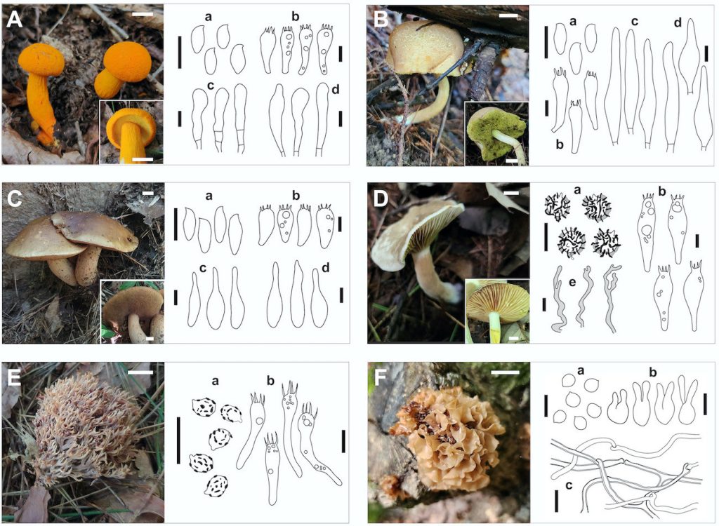
Fig. 1. Basidiocarps and microscopic structures of the six fungal species: (A) Aureoboletus miniatoaurantiacus, (B) Xerocomus galbanus, (C) Boletus bainiugan, (D) Lactarius mirus, (E) Ramaria rubella, and (F) Phaeotremella yunnanensis. Scale bars: basidiocarps = 1 cm; microscopic drawings = 10 µm (A–C: a basidiospores, b basidia, c cheilocystidia, d pleurocystidia; D: a basidiospores, b basidia, e hymenophoral pseudocystidia; E: a basidiospores, b basidia; F: a basidiospores, b phragmobasidia).
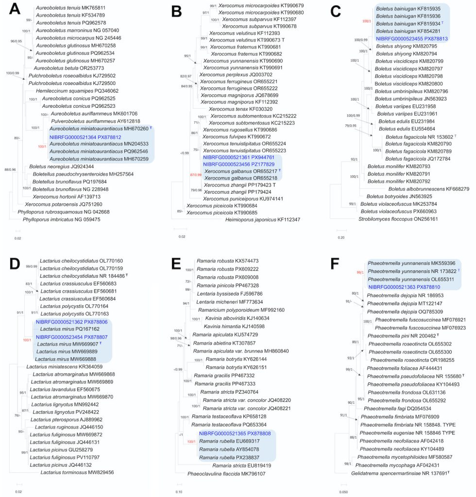
Fig. 2. Maximum likelihood trees of six previously unrecorded species based on different genetic markers. (A–B) Phylogenetic positions of Aureoboletus miniatoaurantiacus, and Xerocomus galbanus inferred from large subunit ribosomal DNA sequences. (C–F) Phylogenetic relationships among Boletus bainiugan, Lactarius mirus, Ramaria rubella, and Phaeotremella yunnanensis based on the internal transcribed spacer region. Numbers above the nodes indicate maximum likelihood bootstrap support (MLBS ≥ 70%, left) and significant Bayesian posterior probability (BPP ≥ 0.95, right). Novel sequences are shown in blue. “T” indicates the type specimens.
Korean name. Ju-Hwang-Saek-Sin-Geu-Mul-Beo-Seot (주황색신그물버섯), proposed here.
The Korean name refers to the orange color of the basidiomata.
Basidiomata: small to medium-sized. Pileus: 18–20 mm in diameter, hemispherical to convex, nearly applanate when mature; surface bright yellow to yellow-orange or reddish-orange, tomentose to pulverulent; context white, unchanged in color when exposed. Hymenophore: tubular, adnate to subdecurrent; pores angular to rounded, 0.5–1 per mm, yellow to olive-yellow, unchanging in color when bruised; tubes 5–10 mm deep. Stipe: 40–50 × 4–6 mm, cylindrical, straight to slightly curved; surface smooth to faintly fibrous with longitudinal streaks, concolorous with pileus; basal mycelium white; context white, unchanged in color when exposed.
Basidiospores: 8–10.5 × 4.5–5.5 µm (Q = 1.7–2.1), ovoid and inequilateral in side view, ovoid in front view, smooth, thin-walled, hyaline to pale yellowish in 5% KOH. Basidia: 29–38 (–40) × 8–12 µm, clavate to subclavate, mostly 4-spored, rarely 2-spored, sterigmata present. Cheilocystidia: abundant, (25–) 32–60 × (7.5–) 8.5–12 µm, clavate to subfusiform or narrowly ventricose, thin-walled, hyaline. Pleurocystidia: (28–) 32–53 (–59) × (6.5–) 8–12 (–13.5) µm, similar to cheilocystidia in shape, mostly ventricose-rostrate to fusiform, thin-walled, scattered.
Habitat: Solitary to scattered in deciduous forests, typically associated with broad-leaved trees (Quercus spp.).
Specimens examined: Korea, Gyeongsangbuk-do, Mungyeong-si, Gaeun-eup, Wanjang-ri (Mt. Songnisan), 36.667534°N, 127.944221°E, alt. 465 m, 10 Sep. 2024, coll. JS Lee, JY Park, and SY Choi, specimen No. NIBRFG0000521364 (JS20240910-11; GenBank accession No. PX878812); Korea, Chungcheongbuk-do, Goesan-gun, Chilseong-myeon, Ssangok-ri (Mt. Songnisan), 36.705627°N, 127.928429°E, alt. 741 m, 5 Aug. 2024, coll. JS Lee, JY Park, and SY Choi, specimen No. NIBRFG0000523453 (JS20240805-06; GenBank accession No. PX878811)
Remarks: Aureoboletus miniatoaurantiacus is characterized by small bright reddish-orange basidiomata and a tomentose or pulverulent pileus. This species is clearly distinguished from the two previously reported Korean species of the genus—A. thibetanus and A. sinobadius—by the distinct shape and coloration of its basidiomata. However, A. auriflammeus, which was originally described in North America, is morphologically similar to A. miniatoaurantiacus. The former is distinguished by a distinctly reticulate stipe and relatively narrow basidiospores (8–12 × 3–5 µm) [16].
Korean name. Gal-Saek-Mang-Geu-Mul-Beo-Seot (갈색망그물버섯), proposed here.
The Korean name refers to the brown color of the basidiomata.
Basidiomata: medium to large-sized. Pileus: 50–120 mm in diameter, convex to broadly convex, applanate when mature; surface dry or glabrous, slightly rugose in some parts, dark brown to chestnut brown (umber), then brown to yellow-brown; context white, unchanged in color when exposed. Hymenophore: tubular, adnate to slightly depressed around the stipe; pores circular to angular, 1–2 per mm, yellow-brown to darker brown, unchanging in color when bruised; tubes 5–8 mm long, concolorous with the pore surface. Stipe: 50–70 × 25–40 mm, central, clavate to subcylindrical, significantly swollen at the base, solid; surface pale cream to brownish-white, distinctly covered with slightly darker brownish reticulations over the entire stipe; context white, unchanged in color when exposed; basal mycelium white.
Basidiospores: 11–14 (–15.4) × 4–5.5 µm (Q = 2.3–3.0), subfusiform-inequilateral in side view with a suprahilar depression, elliptic-fusiform in front view, smooth, hyaline to pale yellowish-brown in 5% KOH. Basidia: 25–33 × 10–12.5 (–13.5) µm, clavate to broadly clavate, mostly 4-spored, sterigmata distinct. Cheilocystidia: 25–42 × 7.5–11 (–12) µm, abundant, fusiform-ventricose with a long neck or mucronate apex, thin-walled, hyaline. Pleurocystidia: 40–50 × 9–12 µm, similar to cheilocystidia in shape, fusiform-ventricose to lageniform, thin-walled, scattered.
Habitat: Solitary or scattered on the ground in a coniferous or mixed forest.
Specimen examined: Korea, Gyeongsangbuk-do, Sangju-si, Hwabuk-myeon, Jangam-ri (Mt. Songnisan), 36.571064°N, 127.884026°E, alt. 446 m, 24 Sep. 2024, coll. JS Lee, JY Park, and SY Choi, specimen No. NIBRFG0000523455 (JS20240924-32; GenBank accession Nos. PX878813 and PX944760).
Remarks: Boletus bainiugan is characterized by a yellowish-brown to brown pileus with a rugose surface and a stipe covered with pale brown reticulations. It is morphologically distinguished from its most closely related congener, Boletus edulis, by its darker, distinctly rugose pileus surface and a more pronounced, brownish-tinged reticulum that frequently extends further down the length of the stipe. It differs from Boletus reticulatus in its subviscid pileus texture when moist [17].
Korean name. Hwang-Nok-Saek-San-Geu-Mul-Beo-Seot (황녹색산그물버섯), proposed here.
The Korean name refers to the characteristic yellow-green hue of the pore surface.
Basidiomata: small- to medium-sized. Pileus: 30–55 mm in diameter, convex at first, becoming broadly convex to applanate; surface dry, finely tomentose to velvety, sometimes cracked when mature; grayish-white, pale yellowish-brown to ochraceous-brown; context white to pale yellow, unchanging in color when exposed. Hymenophore: tubular, adnate to slightly depressed around the stipe; pores angular, 1–2 mm wide in mature specimens; bright lemon-yellow to yellow green, weakly turning blue when bruised; tubes 5–8 mm long, concolorous with the pore surface. Stipe: 40–80 × 5–12 mm, central, cylindrical, or tapering slightly toward the base; surface pale yellow to pale brown, often whitish toward the base, covered with longitudinally oriented reticulum confined to the apex; context light yellow to pale yellow, unchanged in color when exposed; basal mycelium white.
Basidiospores: 12–14.5 × 4.5–5.5 µm (Q = 2.2–2.9), subfusiform, with slight suprahilar depression in side view, subfusoid in face view, smooth, hyaline to pale yellowish in 5% KOH. Basidia: (30–) 32–41 (–43) × 8–12 (–13) µm clavate to narrowly clavate, mostly 4-spored with distinct sterigmata. Cheilocystidia: 45–77 × 9.5–12 µm, abundant, fusiform to narrowly ventricose-rostrate, thin-walled, hyaline. Pleurocystidia: 50–84 × 9–15 µm, similar to cheilocystidia in shape but generally larger and more prominently ventricose, scattered, thin-walled.
Habitat: Solitary or scattered on the ground in a coniferous or mixed forest.
Specimens examined: Korea, Chungcheongbuk-do, Goesan-gun, Chilseong-myeon, Saeun-ri (Mt. Songnisan), 36.715004°N, 127.857452°E, alt. 504 m, 6 Aug. 2024, coll. JS Lee, JY Park, and SY Choi, specimen No. NIBRFG0000521361 (JS20240806-09; GenBank accession Nos. PX878809 and PX944761); Korea, Chungcheongbuk-do, Goesan-gun, Chilseong-myeon, Saeun-ri (Mt. Songnisan), 36.715004°N, 127.857452°E, alt. 504 m, 18 Aug. 2025, coll. JY Park and DG Choi, specimen No. NIBRFG0000523456 (QM20250818-26; GenBank accession Nos. PZ177829 and PZ213864).
Remarks: Xerocomus galbanus is primarily characterized by a distinctive greenish-yellow hymenophore that turns weakly blue when bruised and by a stipe bearing elongated reticula on the upper part. Among the six Xerocomus species recorded in Korea, this species is morphologically most similar to Xerocomus subtomentosus. Both taxa share a yellowish-brown to olive-brown pileus and relatively large yellow tubes that typically exhibit a weak or absent bluing reaction. However, X. galbanus is distinguished from X. subtomentosus by its relatively smaller basidiospores (12–14.5 × 4.5–5.5 µm) and significantly larger, more prominently ventricose pleurocystidia [18].
Korean name. Yeon-Norang-Jeot-Beo-Seot (연노랑젖버섯), proposed here.
The Korean name refers to the pale yellow color of the basidiomata.
Basidiomata: small- to medium-sized. Pileus: 30–50 mm in diameter, convex to plano-convex at first, then applanate with a shallow depression at center or concave when mature; surface smooth, rugose or grooved, dry to velvety, grayish brown to light brown, slightly darker at the center; margin slightly incurved to decurved, becoming wavy with age; context whitish, turning yellow owing to the latex when exposed. Lamellae: decurrent to slightly adnate, distant to moderately crowded, pale cream to light buff, sometimes with small yellowish spots; lamellulae present. Stipe: 40–60 × 6–10 mm, central, cylindrical, slightly tapering downward; surface dry to finely fibrillose, whitish to pale cream; context whitish, turning yellow in color owing to the latex when exposed. Latex: copious, white, immediately changing to yellow upon exposure.
Basidiospores: (7.5–) 8–10 (–10.5) × 7.5–9.5 (–10.5) µm (Q = 1.0–1.1), globose to subglobose, hyaline; ornamentation 1.5–3 µm high, consisting of prominent, irregular ridges and warts forming a partial to nearly complete reticulum. Basidia: 52.5–66 (–71) × 13–16.5 µm, clavate to broadly ventricose-clavate, 4-spored, with granular contents and distinct sterigmata. Pleuromacrocystidia and cheilomacrocystidia: absent. Hymenophoral pseudocystidia: 3–6 µm in diameter, common, cylindrical to tortuous or filamentous, buried in hymenium or rarely projecting, pale brown.
Habitat: Solitary or scattered on soil in a deciduous forest.
Specimens examined: Korea, Chungcheongbuk-do, Goesan-gun, Chilseong-myeon, Saeun-ri (Mt. Songnisan), 36.715012°N, 127.856453°E, alt. 538 m, 6 Aug. 2024, coll. JS Lee, JY Park, and SY Choi, specimen No. NIBRFG0000521362 (JS20240806-04; GenBank accession No. PX878806); Korea, Chungcheongbuk-do, Goesan-gun, Chilseong-myeon, Saeun-ri, 36.754269°N, 127.845154°E, alt. 207 m, 26 Aug. 2024, coll. JS Lee, JY Park, and SY Choi, specimen No. NIBRFG0000523454 (JS20240826-03; GenBank accession No. PX878807).
Remarks: Lactarius mirus is characterized by grayish-brown to light-brown basidiomata, adnate to slightly decurrent lamellae, and copious latex that rapidly changes from white to bright yellow upon exposure. L. mirus resembles species within the chrysorrheus group, such as L. lutescens and L. citrinus, which also possess latex that changes from white to bright yellow upon exposure. However, L. mirus is distinguished by its paler, non-zonate, brownish pileus and the absence of the typical orange to reddish pileus tones found in L. lutescens and L. citrinus [19].
Korean name. Po-Do-Saek-Ssa-Ri-Beo-Seot (포도색싸리버섯), proposed here.
The Korean name refers to the vinaceous or grape-like color of the basidiomata.
Basidiomata: medium-sized. Fruitbody: overall dimensions approximately 40–90 mm in width and 35–60 mm in height, coralloid; branches numerous, highly subdivided, vertically oriented; internodes decreasing in length toward the apices; surface smooth to slightly rugose, pale ochraceous-pink to reddish-brown; apices multifid or dentate, concolorous with branches or slightly paler yellowish in younger growth stages, becoming darker or brownish with maturity or upon bruising. Stipe: relatively short and indistinct; surface whitish to pale tan, occasionally showing white rhizomorphs at the point of substrate attachment. Context: brittle when dry, white to pale vinaceous.
Basidiospores: 5.8–7.0 (–7.5) × 4.0–5.0 µm (Q = 1.0–1.3), globose to subglobose, hyaline to pale yellowish; ornamentation distinct, consisting of numerous small, cyanophilous warts or short ridges scattered over the surface; apiculus prominent and eccentric. Basidia: (33–) 38.5–57 × 7.0–8.3 µm, clavate to narrowly clavate, often slightly curved or flexuous at the base, 4-spored; sterigmata 4–6 µm long, straight to slightly curved; often containing small oil droplets or granular contents.
Habitat: Growing gregariously on leaf litter and decaying wood.
Specimen examined: Korea, Chungcheongbuk-do, Boeun-gun, Songnisan-myeon, Sanae-ri, 36.556709°N, 127.829277°E, alt. 440 m, 24 Sep. 2024, coll. JS Lee, JY Park, and SY Choi, specimen No. NIBRFG0000521365 (JS20240924-24; GenBank accession No. PX878808).
Remarks: Ramaria rubella is most frequently confused with Ramaria stricta because both taxa typically occur on decaying wood and share similar macroscopic structures. However, R. rubella is primarily distinguished from R. stricta by its distinctly reddish to vinaceous branches, relatively shorter and broader ellipsoid basidiospores 5.8–7.0 (–7.5) × 4.0–5.0 µm (Q=1.0–1.3), and a milder taste that lacks the intense bitterness characteristic of the latter [20,21].
Tremellales Fr. 1821
Tremellaceae Fr. 1821
Phaeotremella Rea, Trans. Br. mycol. Soc. 3(5): 377 (1912)
Korean name. Jaet-Bit-Mok-i-Sok (잿빛목이속), proposed here.
The Korean name refers to the grayish-brown (ashy) color of the basidiomata within this genus.
Korean name. Mul-Gyeol-Jaet-Bit-Mok-I (물결잿빛목이) proposed here.
The Korean name refers to the characteristic wavy or ruffled shape of its foliose fruit body.
Basidiomata: medium-sized, approximately 45–65 mm in diameter and 20–35 mm in height, gelatinous when moist, becoming somewhat tougher or brittle when dry, foliose to cerebriform; structure crowded, undulating, and flattened lobes arising from a central attachment point on the substrate; lobes thin, translucent to opaque, with wavy or ruffled margins; surface smooth, shiny, and viscid when wet, pale brownish-orange to cinnamon-brown.
Basidiospores: 7–9 (–9.5) × 6–8 µm (Q = 1.0–1.3), globose to broadly ellipsoid, with a distinct apiculus, smooth, hyaline. Phragmobasidia: 17–22 × 8.5–12.5 µm, globose to subglobose, with longitudinal or cruciate septa, bearing long, cylindrical sterigmata, thin-walled, hyaline. Hyphae: clamped, thin- to slightly thick-walled, 2–3 µm diam, hyaline, inflated, branched and slightly interwoven.
Habitat: Growing on deciduous tree stumps.
Specimen examined: Korea, Gyeongsangbuk-do, Mungyeong-si, Gaeun-eup, Wanjang-ri, 36.668484°N, 127.94249°E, alt. 491 m, 10 Sep. 2024, coll. JS Lee, JY Park, and SY Choi, specimen No. NIBRFG0000521363 (JS20240910-17; GenBank accession No. PX878810).
Remarks: Phaeotremella yunnanensis is characterized by gelatinous lobed fruiting bodies with a distinct brownish coloration, typically occurring on fallen broadleaf branches. Although it macroscopically resembles P. foliacea owing to its brownish tint, P. yunnanensis is distinguished by its relatively narrower basidiospores (as reported in original descriptions, approx. 5.3–9.1 × 4.7–8.5 µm) and the presence of swollen cells that produce ellipsoid to subglobose conidia. In contrast, P. foliacea is characterized by slightly thick-walled basidia and a specific ecological preference for coniferous hosts [22].
This study confirms the occurrence of six macrofungal species previously unrecorded in Korea, expanding our understanding of fungal diversity within the Songnisan National Park (part of the Baekdudaegan mountain range). The integration of morphological characteristics with ITS and LSU rDNA-based phylogenetic analyses provided robust evidence for their identification.
The genus Aureoboletus (Boletaceae, Boletales) is morphologically defined by its viscid to slimy pileus, unchanging golden-yellow hymenophore, and a characteristic slight depression at the stipe apex [16,23]. Recent molecular phylogenetic studies have resolved the position of Aureoboletus within the Boletaceae (subfamily Xerocomoideae), confirming its close phylogenetic relationship with Boletellus, Hemileccinum, Heimioporus, and Xerocomus [16]. To date, only two species belonging to this genus, A. thibetanus and A. sinobadius, have been formally recorded in the Korean fungal flora [2,24]. Aureoboletus miniatoaurantiacus is distinguished by its bright orange-yellow basidiomata, tomentose or pulverulent pileus surface, light orange to orange hymenophore. It differs from the widely distributed A. thibetanus in its smaller basidiocarps and a smooth to finely pruinose stipe, in contrast to the larger yellowish-brown pileus and distinctly rugose to pitted-reticulate stipe of the latter [16,25]. Additionally, A. sinobadius is differentiated from A. miniatoaurantiacus by its brownish-red to vinaceous brown pileus [16,24].
The genus Boletus (Boletaceae, Boletales) is morphologically characterized by robust fleshy basidiocarps, a tubulate hymenophore with small circular pores, and a central stipe that typically features a distinct reticulum [26]. Microscopically, the genus is defined by smooth, subfusiform to ellipsoid, olivaceous-brown spores, and a divergent hymenophoral trama. Although the National List of Species of Korea [2] currently records 30 species under Boletus sensu lato, many of these species are undergoing taxonomic reassessment following recent phylogenetic redefinitions. The identification of Boletus bainiugan is of particular taxonomic significance because of its close morphological affinity with B. edulis [17]. Although B. bainiugan is a well-known edible species traditionally traded in Southwestern China, there has been no formal record of its occurrence in Korea to date. The specimen examined in this study exhibited a pale brown pileus and distinct stipe reticulation, which are consistent with the diagnostic descriptions of the species. Our findings confirm the occurrence of B. bainiugan in Korea and provide a basis for further biogeographical studies on the B. edulis complex in East Asia.
The genus Xerocomus (Boletaceae, Boletales) is traditionally characterized by a relatively slender stature, a dry tomentose to subtomentose pileus, and a hymenophore with wide angular pores that typically exhibit a cyanescent (bluing) reaction upon bruising [26]. In South Korea, the documented diversity of this genus currently comprises six species [2]. Among these, Xerocomus galbanus, a taxon recently described in China, is distinguished by its characteristic yellowish-green coloration [18]. Although the ITS sequence analysis of specimen NIBRFG0000521361 showed high similarity to both X. galbanus and X. zhangii, a comparative morphological assessment strongly supported its affinity to X. galbanus. The microscopic characteristics of our specimen align more closely with the diagnostic ranges described for X. galbanus than those for X. zhangii. Specifically, the basidiospores of specimen NIBRFG0000521361 had the following dimensions—12–14.5 × 4.5–5.5 µm (Q = 2.2–2.9)—which largely overlap with those of X. galbanus—(12–)13–15 × (4–)4.5–6 µm (Q = 2.3–3.0)—while being generally larger and more elongated than those of X. zhangii—(8.5–)9–11.5(–12) × 4–5 µm (Q = 2.0–2.44) [27]. Notably, the Q-range of the present specimen was closer to that of X. galbanus than to that of X. zhangii. Furthermore, the tef-1α sequence of our specimen exhibited a significantly higher similarity to X. galbanus (99.1%) than to X. zhangii (94.5%), providing robust molecular evidence for its taxonomic identity.
The genus Lactarius Pers. (Russulaceae, Russulales) represents one of the most diverse lineages of ectomycorrhizal fungi, distinguished by the exudation of milky or pigmented latex upon injury to the basidiocarp [26]. Overall, 97 species of Lactarius have been documented in South Korea [2]. In the present study, we report Lactarius mirus, a taxon belonging to the section Plinthogalus, characterized by its whitish gray to dark brown pileus and stipe. Although L. mirus shares a strong morphological affinity with L. flavorosescens, it can be clearly distinguished by the spore ornamentation of the latter, which is typically zebroid, lower (< 2.5 µm), and rarely branching [28].
The genus Phaeotremella Rea (Phaeotremellaceae, Tremellales) comprises dimorphic mycoparasitic fungi characterized by foliaceous gelatinous basidiocarps with brownish to reddish-brown hues. Although historically circumscribed within Tremella, recent multi-gene phylogenetic analyses have established Phaeotremella as a distinct genus within the family Tremellaceae [29]. Morphologically, it is defined by globose to subglobose, longitudinally cruciate septate basidia, with a specialized parasitic association with wood-decaying fungi [22,30]. In South Korea, the diversity of Phaeotremella remains understudied compared to that of the major agaricoid lineages. In this study, we report P. yunnanensis for the first time in Korea. This species is morphologically distinguishable from its close congeners, P. foliacea and P. roseotincta, by its distinct brownish foliaceous basidiocarps. Its identification was further supported by significant genetic divergence, as previously established [22].
The genus Ramaria Fr. ex Bonord. (Gomphaceae, Gomphales) represents a morphologically complex lineage of coral fungi characterized by coralloid basidiocarps with repeated branching, positively geotropic branches, and cyanophilous, ornamented basidiospores [26]. While traditional infrageneric classifications are based on these morphological traits, recent molecular phylogenetic evidence has revealed that many traditional subgenera are polyphyletic, necessitating recircumscription of the genus using multi-gene sequence data [31]. In South Korea, Ramaria is a significant component of forest fungal diversity, with 16 species formally documented to date [2]. Among these, R. rubella is readily distinguished by its numerous, dense, and repeatedly polydichotomically branched pinkish-red to vinaceous-brown branches, as well as its taste is less bitter and acrid compared to other species in the same genus [20]. In this study, the ITS sequence of our specimen exhibited 100% similarity with reference strains from China, supporting its taxonomic placement and confirming its distribution across Northeast Asia.
In conclusion, this study provides valuable data to the Korean mycobiota checklist. The addition of these six species increases the known fungal biodiversity of the region and emphasizes the importance of protected areas, such as Songnisan National Park, for maintaining fungal diversity. Continuous efforts in both field collection and taxonomic research are essential to develop a complete inventory of Korean fungi.
This study confirms the occurrence of six macrofungal species previously unrecorded in Korea, expanding our understanding of fungal diversity within the Songnisan National Park (part of the Baekdudaegan mountain range). The integration of morphological characteristics with ITS and LSU rDNA-based phylogenetic analyses provided robust evidence for their identification.
The genus Aureoboletus (Boletaceae, Boletales) is morphologically defined by its viscid to slimy pileus, unchanging golden-yellow hymenophore, and a characteristic slight depression at the stipe apex [16,23]. Recent molecular phylogenetic studies have resolved the position of Aureoboletus within the Boletaceae (subfamily Xerocomoideae), confirming its close phylogenetic relationship with Boletellus, Hemileccinum, Heimioporus, and Xerocomus [16]. To date, only two species belonging to this genus, A. thibetanus and A. sinobadius, have been formally recorded in the Korean fungal flora [2,24]. Aureoboletus miniatoaurantiacus is distinguished by its bright orange-yellow basidiomata, tomentose or pulverulent pileus surface, light orange to orange hymenophore. It differs from the widely distributed A. thibetanus in its smaller basidiocarps and a smooth to finely pruinose stipe, in contrast to the larger yellowish-brown pileus and distinctly rugose to pitted-reticulate stipe of the latter [16,25]. Additionally, A. sinobadius is differentiated from A. miniatoaurantiacus by its brownish-red to vinaceous brown pileus [16,24].
The genus Boletus (Boletaceae, Boletales) is morphologically characterized by robust fleshy basidiocarps, a tubulate hymenophore with small circular pores, and a central stipe that typically features a distinct reticulum [26]. Microscopically, the genus is defined by smooth, subfusiform to ellipsoid, olivaceous-brown spores, and a divergent hymenophoral trama. Although the National List of Species of Korea [2] currently records 30 species under Boletus sensu lato, many of these species are undergoing taxonomic reassessment following recent phylogenetic redefinitions. The identification of Boletus bainiugan is of particular taxonomic significance because of its close morphological affinity with B. edulis [17]. Although B. bainiugan is a well-known edible species traditionally traded in Southwestern China, there has been no formal record of its occurrence in Korea to date. The specimen examined in this study exhibited a pale brown pileus and distinct stipe reticulation, which are consistent with the diagnostic descriptions of the species. Our findings confirm the occurrence of B. bainiugan in Korea and provide a basis for further biogeographical studies on the B. edulis complex in East Asia.
The genus Xerocomus (Boletaceae, Boletales) is traditionally characterized by a relatively slender stature, a dry tomentose to subtomentose pileus, and a hymenophore with wide angular pores that typically exhibit a cyanescent (bluing) reaction upon bruising [26]. In South Korea, the documented diversity of this genus currently comprises six species [2]. Among these, Xerocomus galbanus, a taxon recently described in China, is distinguished by its characteristic yellowish-green coloration [18]. Although the ITS sequence analysis of specimen NIBRFG0000521361 showed high similarity to both X. galbanus and X. zhangii, a comparative morphological assessment strongly supported its affinity to X. galbanus. The microscopic characteristics of our specimen align more closely with the diagnostic ranges described for X. galbanus than those for X. zhangii. Specifically, the basidiospores of specimen NIBRFG0000521361 had the following dimensions—12–14.5 × 4.5–5.5 µm (Q = 2.2–2.9)—which largely overlap with those of X. galbanus—(12–)13–15 × (4–)4.5–6 µm (Q = 2.3–3.0)—while being generally larger and more elongated than those of X. zhangii—(8.5–)9–11.5(–12) × 4–5 µm (Q = 2.0–2.44) [27]. Notably, the Q-range of the present specimen was closer to that of X. galbanus than to that of X. zhangii. Furthermore, the tef-1α sequence of our specimen exhibited a significantly higher similarity to X. galbanus (99.1%) than to X. zhangii (94.5%), providing robust molecular evidence for its taxonomic identity.
The genus Lactarius Pers. (Russulaceae, Russulales) represents one of the most diverse lineages of ectomycorrhizal fungi, distinguished by the exudation of milky or pigmented latex upon injury to the basidiocarp [26]. Overall, 97 species of Lactarius have been documented in South Korea [2]. In the present study, we report Lactarius mirus, a taxon belonging to the section Plinthogalus, characterized by its whitish gray to dark brown pileus and stipe. Although L. mirus shares a strong morphological affinity with L. flavorosescens, it can be clearly distinguished by the spore ornamentation of the latter, which is typically zebroid, lower (< 2.5 µm), and rarely branching [28].
The genus Phaeotremella Rea (Phaeotremellaceae, Tremellales) comprises dimorphic mycoparasitic fungi characterized by foliaceous gelatinous basidiocarps with brownish to reddish-brown hues. Although historically circumscribed within Tremella, recent multi-gene phylogenetic analyses have established Phaeotremella as a distinct genus within the family Tremellaceae [29]. Morphologically, it is defined by globose to subglobose, longitudinally cruciate septate basidia, with a specialized parasitic association with wood-decaying fungi [22,30]. In South Korea, the diversity of Phaeotremella remains understudied compared to that of the major agaricoid lineages. In this study, we report P. yunnanensis for the first time in Korea. This species is morphologically distinguishable from its close congeners, P. foliacea and P. roseotincta, by its distinct brownish foliaceous basidiocarps. Its identification was further supported by significant genetic divergence, as previously established [22].
The genus Ramaria Fr. ex Bonord. (Gomphaceae, Gomphales) represents a morphologically complex lineage of coral fungi characterized by coralloid basidiocarps with repeated branching, positively geotropic branches, and cyanophilous, ornamented basidiospores [26]. While traditional infrageneric classifications are based on these morphological traits, recent molecular phylogenetic evidence has revealed that many traditional subgenera are polyphyletic, necessitating recircumscription of the genus using multi-gene sequence data [31]. In South Korea, Ramaria is a significant component of forest fungal diversity, with 16 species formally documented to date [2]. Among these, R. rubella is readily distinguished by its numerous, dense, and repeatedly polydichotomically branched pinkish-red to vinaceous-brown branches, as well as its taste is less bitter and acrid compared to other species in the same genus [20]. In this study, the ITS sequence of our specimen exhibited 100% similarity with reference strains from China, supporting its taxonomic placement and confirming its distribution across Northeast Asia.
In conclusion, this study provides valuable data to the Korean mycobiota checklist. The addition of these six species increases the known fungal biodiversity of the region and emphasizes the importance of protected areas, such as Songnisan National Park, for maintaining fungal diversity. Continuous efforts in both field collection and taxonomic research are essential to develop a complete inventory of Korean fungi.
No potential conflicts of interest relevant to this article were reported.
This study was supported by grants from the National Institute of Biological Resources (NIBR) and the Honam National Institute of Biological Resources (HNIBR) funded by the Ministry of Environment (MOE) of the Republic of Korea (NIBR202502103, HNIBR202101111). We also thank the Korea National Park Research Institute (KNPRI) for their support.
1. Kirk PM, Cannon PF, Minter DW, Stalpers JA. Ainsworth & Bisby’s dictionary of the fungi. 10th ed. Wallingford: CABI; 2008.
[DOI]
2. National Institute of Biological Resources. National list of species of Korea [Internet]. Incheon: NIBR; 2025 [cited 2026 Apr 12]. Available from https://species.nibr.go.kr/national-list
3. Park YW, Koo CD, Lee HY, Ryu SR, Kim TH, Cho YG. Relationship between macrofungal fruiting and environmental factors in the Songnisan National Park. Korean J Environ Ecol 2010;24(6):657-79.
4. Korea National Park Service. Introduction to Songnisan National Park [Internet]. Wonju: Korea National Park Service; 2024 [cited 2026 Apr 12]. Available from https://www.knps.or.kr
5. Cho DH, Ryu CI. The mycoflora of higher fungi in Mt. Songni. Rep Korean Assoc Conserv Nat 1991;29:237-45.
6. Lee JS, Kim CM, Park JY, Ryoo KH, Kim KM, Yoon YG, Jung HS. 2004. Unrecorded higher fungi in Songnisan National Park. Mycobiology 2004;32(2):68-73. https://doi.org/10.4489/MYCO.2004.32.2.068
[DOI]
7. Jung HS. Natural resources survey of Songnisan National Park-14. Higher fungi: Higher fungi in the southern region. Wonju: Korea National Park Service; 2003. p. 487-518.
8. Vilgalys R, Hester M. Rapid genetic identification and mapping of enzymatically amplified rDNA from several Cryptococcus species. J Bacteriol 172(8):4238-46. https://doi.org/10.1128/jb.172.8.4238-4246.1990
[DOI][PubMed][PMC]
9. Gardes M, Bruns TD. ITS primers with enhanced specificity for basidiomycetes: Application to the identification of mycorrhizae and rust. Mol Ecol 1993;2:113-8. https://doi.org/10.1111/j.1365-294x.1993.tb00005.x
[DOI][PubMed]
10. White TJ, Bruns TD, Lee SB, Taylor JW. Amplification and direct sequencing of fungal ribosomal RNA genes for phylogenetics. In: Innis MA, Gelfand DH, Sninsky JJ, White TJ, editors. PCR protocols: A guide to methods and applications. San Diego: Academic Press; 1990. p. 315-22. https://doi.org/10.1016/B978-0-12-372180-8.50042-1
[DOI][PubMed][PMC]
11. Rehner SA, Buckley E. A Beauveria phylogeny inferred from nuclear ITS and EF1-α sequences: Evidence for cryptic diversification and links to Cordyceps teleomorphs. Mycologia 2005;97(1):84-98. https://doi.org/10.3852/mycologia.97.1.84
[DOI][PubMed][PMC]
12. Tamura K, Stecher G, Kumar S. MEGA11: Molecular evolutionary genetics analysis version 11. Mol Biol Evol 2021;38(7):3022-7. https://doi.org/10.1093/molbev/msab120
[DOI][PubMed][PMC]
13. Katoh K, Standley DM. MAFFT multiple sequence alignment software, version 7: Improvements in performance and usability. Mol Biol Evol 2013;30:772-80. https://doi.org/10.1093/molbev/mst010
[DOI][PubMed][PMC]
14. Darriba D, Taboada GL, Doallo R, Posada D. jModelTest 2: More models, new heuristics and parallel computing. Nat Methods 2012;9:772. https://doi.org/10.1038/nmeth.2109
[DOI][PubMed][PMC]
15. Ronquist F, Huelsenbeck JP. MrBayes 3: Bayesian phylogenetic inference under mixed models. Bioinformatics 2003;19:1572-4. https://doi.org/10.1093/bioinformatics/btg180
[DOI][PubMed]
16. Zhang M, Li TH, Wang CQ, Zeng NK, Deng WQ. Phylogenetic overview of Aureoboletus (Boletaceae and Boletales) with descriptions of six new species from China. MycoKeys 2019;61:111-45. https://doi.org/10.3897/mycokeys.61.47520
[DOI][PubMed][PMC]
17. Dentinger BTM. Boletus bainiugan Dentinger sp. nov. Index Fungorum 2013;29:1.
18. Mao N, Zhao TY, Zhang YX, Li T, Lv JC, Fan L. Boletaceae from Shanxi Province, northern China, with the descriptions of ten new species. Mycosphere 2023;14(1):2013-91. https://doi.org/10.5943/mycosphere/14/1/24
[DOI]
19. Lee H, Wissitrassameewong K, Park MS, Verbeken A, Eimes J, Lim YW. Taxonomic revision of the genus Lactarius (Russulales, Basidiomycota) in Korea. Fungal Divers 2019;95:275-335. https://doi.org/10.1007/s13225-019-00425-6
[DOI]
20. Łuszczyński J. Ramaria rubella (Schaeff.) R. H. Petersen – the first record in Central and Eastern Europe. Acta Soc Bot Pol 2008;77(3):241-3. https://doi.org/10.5586/asbp.2008.030
[DOI]
21. Petersen RH. Contribution toward a monograph of Ramaria. I. some classic species redescribed. Am J Bot 1974;61(7):739-48. https://doi.org/10.1002/j.1537-2197.1974.tb12296.x
[DOI]
22. Yuan HS, Lu X, Dai YC, Hyde KD, Kan YH, Kušan I, He SH, Liu NG, Sarma VV, Zhao CL, et al. Fungal diversity notes 1277-1386: Taxonomic and phylogenetic contributions to fungal taxa. Fungal Divers 2020;104:1-266. https://doi.org/10.1007/s13225-020-00461-7
[DOI]
23. Zhou F, Zhang J, Liu S, Liu Q, Liu C, Luo X, Luo X, Wang L, Sun D, Hua R. Aureoboletus pseudorussellii sp. nov., a newly discovered species in genus Aureoboletus of Zhejiang Province, China, Mycobiology 2025;53(3):305-13. https://doi.org/10.1080/12298093.2025.2480406
[DOI][PubMed][PMC]
24. Gelardi M. Contribution to the knowledge of Chinese boletes. II. Aureoboletus thibetanus s. l., Neoboletus brunneissimus, Pulveroboletus macrosporus, Retiboletus kauffmanii (Part I). Riv Micol Romana 2017;102(3):13-30.
25. Park JY, Lee JS, Kim M, Kim C, Kim NK. Six new recorded boletes from Mudeungsan National Park in Korea. Kor J Mycol 2023;51(4):461-70. http://doi.org/10.4489/kjm.20230047
26. Singer R. The Agaricales in modern taxonomy. 4th ed. Königstein: Koeltz Scientific Books; 1986. p. 977.
27. Xue R, Su LJ, Yu TJ, Xu C, Huang HY, Zeng NK, Zhang GL, Tang LP. Four new species and a new combination of Boletaceae (Boletales) from subtropical and tropical China. J Fungi 2024;10(5):348. https://doi.org/10.3390/jof10050348
[DOI][PubMed][PMC]
28. Wang Z, Wang XH, Qin WQ, Chen ZH, Deng WQ, Zhang JH, Bau T. Lactarius mirus sp. nov. (Russulaceae, Russulales), the first species of Lactarius subg. Plinthogalus with yellowing latex in China. Mycosystema 2021;40(7):1591-604. https://doi.org/10.13346/j.mycosystema.210083
29. Liu XZ, Wang QM, Göker M, Groenewald M, Kachalkin AV, Lumbsch HT, Millanes AM, Wedin M, Yurkov AM, Boekhout T, et al. Towards an integrated phylogenetic classification of Tremellomycetes. Stud Mycol 2015;81:85-147. https://doi.org/10.1016/j.simyco.2015.12.001
[DOI][PubMed][PMC]
30. Spirin V, Malysheva V, Yurkov A, Miettinen O, Larsson KH. Studies in the Phaeotremella foliacea group (Tremellomycetes, Basidiomycota). Mycol Progress 2018;17:451-66. https://doi.org/10.1007/s11557-017-1371-4
[DOI]
31. Giachini AJ, Hosaka K, Nouhra E, Spatafora J, Trappe JM. Phylogenetic relationships of Gomphales based on nuc-25S-rDNA, mit-12S-rDNA, and mit-atp6-DNA combined sequences. Fungal Biol 2010;114(2-3):224-34. https://doi.org/10.1016/j.funbio.2010.01.002
[DOI][PubMed]