
Che-Yeon Park1, Seong-Keun Lim1, Seo-Ryeong Lee1, Hae-Dam Kim1, Seung-Yeol Lee1,2,*, and Hee-Young Jung1,2
1Department of Plant Medicine, Kyungpook National University, Daegu 41566, Korea
2Institute of Plant Medicine, Kyungpook National University, Daegu 41566, Korea
*Correspondence to leesy1123@knu.ac.kr
Korean Journal of Mycology (Kor J Mycol) 2026 June, Volume 54, Issue 2, pages 165-172.
https://doi.org/10.4489/kjm.2026.54.2.6
Received on March 09, 2026, Revised on June 02, 2026, Accepted on June 08, 2026, Published on June 30, 2026.
Copyright © The Korean Society of Mycology.
This is an Open Access article which is freely available under the Creative Commons Attribution-Non-Commercial 4.0 International License (CC BY-NC) (https://creativecommons.org/licenses/by-nc/4.0/).
Acaromyces ingoldii, Morphology, Phylogenetic analyses, Yeast-like fungi
Yeast-like fungi are so named because they produce yeast-like daughter cells, called blastoconidia, by budding from the mycelium [1]. These fungi are phylogenetically related to filamentous ascomycete taxa rather than to true yeasts [2]. Yeast-like fungi belong to various taxonomic groups, including genera belonging to Ascomycota, such as Ascoidea and Cephaloascus or genera belonging to Basidiomycota, such as Inopinatum, Meira, and Acaromyces [3–5]. Exobasidiales, which include yeast-like fungi belonging to the Basidiomycota, are mostly plant-associated fungi, including phytopathogenic, endophytic, and epiphytic species, some of which interact with insects [6]. Despite their wide distribution and morphological and physiological diversity, our understanding of yeast-like fungi remains limited [1]. The order Exobasidiales includes several families, such as Cryptobasidiaceae and Exobasidiaceae; several species within these families have been reported to form galls on the stems or trunks of host plants [7–10]. Cryptobasidiaceae, which occurs mainly in the Lauraceae, is characterized by internal sporulation, forming elongated gastroid basidia inside the host tissues [7,10]. The genus Acaromyces, classified within the order Exobasidiales and family Cryptobasidiaceae, is regarded as a yeast-like fungus that produces yeast-like daughter cells known as blastoconidia [1,5]. Following recent taxonomic revisions, Acaromyces currently comprises only one valid species: A. ingoldii. Based on previous studies, A. ingoldii has been reported from a variety of habitats worldwide, ranging from mite cadavers in the coastal plain of Israel to marine sediments in the South China Sea [11–13]. Historically, species of the genus Acaromyces have been recognized for their pathogenicity toward a wide range of mite species [14]. Until recently, this species had not been reported in Korea. The present study aimed to clarify the taxonomic status of previously unrecorded species in Korea through detailed morphological examinations and phylogenetic analyses.
Healthy apple leaf samples were collected from Miryang-si, Gyeongnam province, Korea (35°35’39.6″N, 128°57’1.8″E). To isolate fungi from the leaf surface, apple leaves were sterilized in 70% ethanol for 1 min and in a 2% sodium hypochlorite solution for 1 min. The samples were placed in double-distilled water (DDW) and washed three times for 30 s each. The sterilized samples were dried on a filter paper for 30 min. Then 100 μL of DDW was added to the surface and mixed thoroughly. The suspension was then spread onto potato dextrose agar (PDA; Difco, Detroit, MI, USA) plates and incubated for 2–3 days at 25℃. Single colonies were then transferred to fresh PDA plates and incubated at 25℃. The isolated fungi were designated KNUF-25-GA222 and selected for molecular, cultural, and morphological analyses.
The strain was cultured at 25°C on PDA and yeast extract peptone glucose agar (YPGA; yeast extract, 10 g; peptone, 5 g; glucose, 40 g; agar, 15 g; and distilled H2O, 1,000 mL) for morphological and cultural characterization. Cultures were incubated for 7 and 21 days in the dark, and characteristics such as the size, color, and shape of the mycelium, and details of the colonies were observed. The cultures were identified by visual observation and examination under a light microscope (BX-50; Olympus, Tokyo, Japan).
For phylogenetic analysis, genomic DNA was extracted from cultures grown on PDA plates using the HiGeneTM Genomic DNA Prep Kit (Biofact, Daejeon, Korea), according to the manufacturer’s instructions. Phylogenetic analysis was conducted based on internal transcribed spacer (ITS) regions and 28S rRNA gene (LSU), which were amplified using the primer pairs ITS1F/ITS4 and LR0R/LR5, respectively [15–17]. The amplified products were purified using ExoSAP-IT (Thermo Fisher Scientific, Waltham, MA, USA) and submitted to Macrogen (Seoul, Korea) for sequencing. The sequences of the obtained strains were deposited in the National Center for Biotechnology Information (NCBI) GenBank database (Table 1).
Table 1. GenBank accession numbers of strains used in this study
| Species | Strain numbers | GenBank accession No. | |
|---|---|---|---|
| ITS | LSU | ||
| Acaromyces ingoldii | KNUF-25-GA222 | LC901949 | LC901950 |
| Acaromyces ingoldii | CBS 10536 | AM991023 | AM991022 |
| Acaromyces ingoldii | CBS 110050T | AY158671 | AY158665 |
| Clinoconidium onumae | HM16-730C | LC522970 | LC523831 |
| Erythrobasidium hasegawianum | JCM 1545T | NR_111008 | AF131058 |
| Exobasidium gracile | DSM 4460 | DQ663700 | DQ663699 |
| Exobasidium rhododendri | CBS 101457 | DQ667153 | DQ667151 |
| Exobasidium vaccinii | TUB 019109 | AB180362 | FJ644526 |
| Laurobasidium hachijoense | MAFF238665 | AB180359 | AB177562 |
| Laurobasidium lauri | M.P. 2371 | MZ159755 | AF487403 |
| Meira argovae | AS006 | AY158676 | AY158670 |
| Meira argovae | CBS 110053T | AY158675 | AY158669 |
| Meira geulakonigii | PM1 | GQ917049 | GQ917048 |
| Meira geulakonigii | CBS 110052T | AY158674 | AY158668 |
| Meira miltonrushii | MCA 3882T | NR_120190 | JX432962 |
| Meira nashicola | CBS 117161T | AB185159 | AB185157 |
Ttype strain. ITS: internal transcribed spacer regions; LSU: 28S rRNA gene.
The strains identified in this study are indicated in bold.
The sequence of strain KNUF-25-GA222 was analyzed for similarity against datasets in the NCBI database using the Basic Local Alignment Search Tool (BLAST). Several related sequences were retrieved from the database for phylogenetic analysis. Phylogenetic trees were constructed using the concatenated sequences of the ITS regions and the LSU gene, employing the maximum-likelihood (ML) method with Molecular Evolutionary Genetics Analysis (MEGA) 11.0 software [18]. An evolutionary distance matrix analysis was performed using the Tamura–Nei model, and bootstrap values were based on 1,000 replications [19].
Acaromyces ingoldii Boekhout, Scorzetti, Gerson & Sztejnb. ex Denchev & T. Denchev, Mycobiota 11:5 (2021) [MB#558279]
Cultures on PDA after 7 days at 25°C, measured 12 mm in diameter. The colonies were tough, pale yellow, velvety, and pruinose with a ridged lamelliform surface, flat marginal zone, and an eroded outermost margin. After 21 days, the colonies grew 24 mm in diameter, appeared greyish brown, were covered with white velvety patches, and the reverse side of the colony was brown (Fig. 1A). Under the same conditions, the colonies were cultured on YPGA for 7 days and were 14 mm in diameter, firm and ridged, whitish, velvety surface, pulvinoid, and furrowed, with the margin eroded. After 21 days, the colonies were 23 mm in diameter, the surface was pruinose and cerebriform, and the reverse side of the colony was yellowish-brown (Fig. 1B). The production of blastoconidia within the mycelium was clearly observed (Fig. 1C). These blastoconidia originated from sterigma-like structures measuring 13–61 × 2–5 μm in size (Fig. 1D and E). Blastoconidia were formed acropetally in chains, became smaller toward the apex of the conidial chain, and were usually formed near the septa of narrow hyaline hyphae (Fig. 1F) [5,20,21]. The strains Acaromyces ingoldii KNUF-25-GA222 and A. ingoldii CBS 110050T exhibited similar cultural characteristics on PDA and YPGA media and shared morphological traits such as velvety pruinose and furrowed colonies, and blastoconidia formed from lateral sterigma-like structures (Table 2).
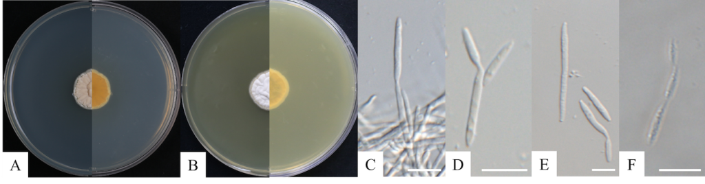
Fig. 1. Cultural and morphological characteristics of Acaromyces ingoldii KNUF-25-GA222. A, B: obverse and reverse view of the colony at 25°C after 21 days on potato dextrose agar (PDA) and yeast extract peptone glucose agar (YPGA), respectively; C: blastoconidia arising from the hyphae; D, E: blastoconidia, F: blastoconidia formed in chains. Scale bars = 10 μm.
Table 2. Cultural and morphological characteristics of the isolated strain KNUF-25-GA222 with reference to Acaromyces species
| Characteristics | Acaromyces ingoldii KNUF-25-GA222a | Acaromyces ingoldii CBS 110050Tb |
|---|---|---|
| Colony on PDA | 24 mm diam, after 21 days at 25°C in the dark velvety pruinose, ridged, furrowed, pale yellow at first, becoming greyish brown | 25 mm diam, after 21 days at 25°C in the dark velvety pruinose, pulvinate, furrowed, whitish at first, becoming greyish brown |
| Colony on YPGA | 23 mm diam, after 21 days at 25°C in the dark; velvety, thin pruinose, pulvinoid, furrowed, margin eroded, whitish | 16 mm diam, after 21 days at 25°C in the dark; velvety, thin pruinose, pulvinoid, furrowed, margin eroded, whitish |
| Blastoconidia | 13–61 × 2–5 µm; sterigma-like, fusiform, aseptate, hyaline | 20–35 × 2–3 µm; sterigma-like, fusiform, shorter near the apex of the chain, hyaline |
afungal strain used in this study; bsource of descriptions [4]; Ttype strain.
PDA: potato dextrose agar; YPGA: yeast extract peptone glucose agar.
Amplification of the ITS region and the LSU gene for the identification of the isolated fungus KNUF-25-GA222 yielded sequences of 556 and 806 bp, respectively. The ITS region exhibited 99.6% similarity with Acaromyces ingoldii CBS 10536 and 98.4% similarity with Laurobasidium hachijoense MAFF238330. In the case of the LSU gene sequence, the strain exhibited 100% similarity to strain A. ingoldii CBS 10536 and 99.9% similarity to L. hachijoense MAFF238665. The ML phylogenetic tree was generated using concatenated sequences of the ITS regions and the LSU gene; strain KNUF-25-GA222 clustered together with A. ingoldii CBS 10536. Thus, based on the morphological and phylogenetic analyses, strain KNUF-25-GA222 was identified as A. ingoldii (Fig. 2).
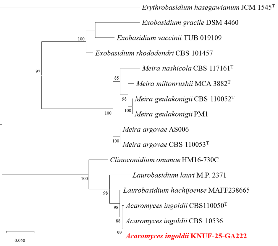
Fig. 2. Maximum-likelihood phylogenetic tree based on a combined dataset of partial sequences of internal transcribed spacer (ITS) regions and 28S rRNA gene (LSU) showing the phylogenetic position of the strain KNUF-25-GA222 among Exobasidiales. Bootstrap values greater than 80% (percentage of 1,000 replications) are shown at branching points. The strain isolated in this study is in bold and red. The tree was rooted using Erythrobasidium hasegawianum JCM 1545T as an outgroup. Bar = 0.050 substitutions per nucleotide position.
The class Exobasidiomycetes comprises seven genera of yeasts and yeast-like fungi, including Acaromyces and Meira. Since protein-coding genes, as well as small subunit rRNA gene and ITS region, are not available at present, only LSU gene sequences have been used to analyze the phylogenetic relationships of yeast species with teleomorphic species in Exobasidiomycetes [22]. Members of the genus Acaromyces are yeast-like fungi belonging to the Exobasidiales order of Exobasidiomycetes and are rarely recorded worldwide [23]. Previous studies have described A. ingoldii as a yeast-like basidiomycete that produces blastoconidia, and the morphological and molecular characteristics observed in the Korean isolate were consistent with those of previously reported A. ingoldii strains [5]. To date, two species belonging to the genus Acaromyces have been documented worldwide. A. laviae was originally described as a yeast-like organism associated with the death of Acarapis woodi, a mite responsible for acarine disease in honeybees [14]. This association suggests the potential pathogenicity of A. laviae in mites. However, since A. laviae has not been formally described, it is considered invalid, whereas A. ingoldii is the only validly reported species. A. ingoldii is a well-known entomopathogenic fungus that exhibits pathogenicity against phytophagous mites [11]. Other studies have revealed that this species was first described based on a culture isolated from the citrus rust mite infesting grapefruit leaves (Citrus paradisi) and was subsequently assayed against several citrus mite species, Eutetranychus orientalis, Tetranychus urticae, Phyllocoptruta oleivora, and Panonychus citri [5,11,12]. Because the pathogenicity of this species against mites has been demonstrated in previous studies, further studies will be conducted to apply this strain to citrus rust mites and other pest mite species to confirm mortality and evaluate its mite-inhibitory activity. In addition, A. ingoldii produces secondary metabolites that inhibit Raffaelea lauricola, the causal agent of laurel wilt, which is an important disease that affects members of the Lauraceae family, including sassafras (Sassafras albidum). In addition, A. ingoldii suppresses the growth of wood-decaying fungi, including brown and white rot species [24,25]. However, related species of A. ingoldii, including Laurobasidium hachijoense, Clinoconidium onumae, and Laurobasidium lauri have been reported to produce gall structures [8–10]. This suggests that A. ingoldii may also be capable of forming galls on hosts of the Lauraceae family, warranting further investigation into the pathogenicity, host specificity, and ecological roles of A. ingoldii. These findings indicate that A. ingoldii deserves further investigation as a potentially novel plant pathogenic fungus, as a biocontrol agent against phytophagous mites, and as a source of useful secondary metabolites. This study is the first report of A. ingoldii in Korea, expands the known geographical distribution of this species, and provides additional taxonomic insights into Acaromyces.
This work was carried out with the support of “Research Program for Agriculture Science and Technology Development (Project No. RS-2025-02305681)” Rural Development Administration, Republic of Korea.
No conflict of interest was reported or declared by the authors.
1. Skinner CE. The yeast-like fungi: Candida and Brettanomyces. Bacteriol Rev 1947;11:227-74. https://doi.org/10.1128/br.11.4.227-274.1947
[DOI][PubMed][PMC]
2. Groenewald M, Hittinger CT, Bensch K, Opulente DA, Shen XX, Li Y, Liu C, LaBella AL, Zhou X, Limtong S, et al. A genome-informed higher rank classification of the biotechnologically important fungal subphylum Saccharomycotina. Stud Mycol 2023;105:1-22. https://doi.org/10.3114/sim.2023.105.01
[DOI][PubMed][PMC]
3. Kurtzman CP. Molecular taxonomy of the yeasts. Yeast 1994;10:1727-40. https://doi.org/10.1002/yea.320101306
[DOI][PubMed]
4. Haelewaters D, Peterson RA, Nevalainen H, Aime MC. Inopinatum lactosum gen. & comb. nov., the first yeast-like fungus in Leotiomycetes. Int J Syst Evol Microbiol 2021;71:004862. https://doi.org/10.1099/ijsem.0.004862
[DOI][PubMed]
5. Boekhout T, Theelen B, Houbraken J, Robert V, Scorzetti G, Gafni A, Gerson U, Sztejnberg A. Novel anamorphic mite-associated fungi belonging to the Ustilaginomycetes: Meira geulakonigii gen. nov., sp. nov., Meira argovae sp. nov. and Acaromyces ingoldii gen. nov., sp. nov. Int J Syst Evol Microbiol 2003;53:1655-64. https://doi.org/10.1099/ijs.0.02434-0
[DOI][PubMed]
6. Cao B, Haelewaters D, Schoutteten N, Begerow D, Boekhout T, Giachini AJ, Gorjón SP, Gunde-Cimerman N, Hyde KD, Kemler M, et al. Delimiting species in Basidiomycota: a review. Fungal Divers 2021;109:181-237. https://doi.org/10.1007/s13225-021-00479-5
[DOI]
7. Begerow D, Bauer R, Oberwinkler F. The Exobasidiales: An evolutionary hypothesis. Mycol Prog 2002;1:187-99. https://doi.org/10.1007/s11557-006-0018-7
[DOI]
8. Shibata S, Ono T, Kobayashi Y, Miyagawa I, Mukai A, Ichioka Y, Takenaka I, Nakano H, Hirooka Y. First report of horn gall disease of Cinnamomum pseudopedunculatum caused by Laurobasidium hachijoense in the Bonin Islands, Japan. J Gen Plant Pathol 2021;87:322-5. https://doi.org/10.1007/s10327-021-01005-w
[DOI]
9. Kakishima M, Nagao H, Ji JX, Sun Y, Denchev CM. Clinoconidium onumae, comb. nov. (Cryptobasidiaceae) producing galls on shoot buds of Cinnamomum tenuifolium in Japan. Phytotaxa 2017;313:175-84. https://doi.org/10.11646/phytotaxa.313.2.3
[DOI]
10. Palfner G. Austrobasidium, a new gall-forming genus of Exobasidiaceae (Exobasidiales, Basidiomycota) on Hydrangea serratifolia from Chile. Aust Syst Bot 2006;19:431-6. https://doi.org/10.1071/SB05026
[DOI]
11. Gerson U, Gafni A, Paz Z, Sztejnberg A. A tale of three acaropathogenic fungi in Israel: Hirsutella, Meira and Acaromyces. Exp Appl Acarol 2008;46:183-94. https://doi.org/10.1007/s10493-008-9202-6
[DOI][PubMed]
12. Paz Z, Gerson U, Sztejnberg A. Assaying three new fungi against citrus mites in the laboratory, and a field trial. BioControl 2007;52:855-62. https://doi.org/10.1007/s10526-006-9060-2
[DOI]
13. Gao XW, Liu HX, Sun ZH, Chen YC, Tan YZ, Zhang WM. Secondary metabolites from the deep-sea derived fungus Acaromyces ingoldii FS121. Molecules 2016;21:371. https://doi.org/10.3390/molecules21040371
[DOI][PMC]
14. Steinhaus EA. Microbial diseases of insects. Annu Rev Microbiol 1957;11:165-82. https://doi.org/10.1146/annurev.mi.11.100157.001121
[DOI][PubMed]
15. White TJ, Bruns T, Lee S, Taylor JW. Amplification and direct sequencing of fungal ribosomal RNA genes for phylogenetics. In: Innis MA, Gelfand DH, Sninsky JJ, White TJ, editors. PCR protocols: A guide to methods and applications. New York: Academic Press; 1990. p. 315-22. https://doi.org/10.1016/B978-0-12-372180-8.50042-1
[DOI][PubMed][PMC]
16. Gardes M, Bruns TD. ITS primers with enhanced specificity for basidiomycetes -application to the identification of mycorrhizae and rusts. Mol Ecol 1993;2:113-8. https://doi.org/10.1111/j.1365-294X.1993.tb00005.x
[DOI][PubMed]
17. Vilgalys R, Hester M. Rapid genetic identification and mapping of enzymatically amplified ribosomal DNA from several Cryptococcus species. J Bacteriol 1990;172:4238-46. https://doi.org/10.1128/jb.172.8.4238-4246.1990
[DOI][PubMed][PMC]
18. Tamura K, Stecher G, Kumar S. MEGA11: Molecular evolutionary genetics analysis version 11. Mol Biol Evol 2021;38:3022-7. https://doi.org/10.1093/molbev/msab120
[DOI][PubMed][PMC]
19. Tamura K, Nei M. Estimation of the number of nucleotide substitutions in the control region of mitochondrial DNA in humans and chimpanzees. Mol Biol Evol 1993;10:512-26. https://doi.org/10.1093/oxfordjournals.molbev.a040023
[DOI]
20. Boekhout T. Acaromyces Boekhout, Scorzetti, Gerson & Sztejnberg (2003). In: Kurtzman CP, Fell JW, Boekhout T, editors. The yeasts: A taxonomic study. 5th ed. Amsterdam: Elsevier; 2011. p. 1603-5. https://doi.org/10.1016/B978-0-444-52149-1.00135-X
[DOI]
21. Yasuda F, Yamagishi D, Akamatsu H, Izawa H, Kodama M, Otani H. Meira nashicola sp. nov., a novel basidiomycetous, anamorphic yeastlike fungus isolated from Japanese pear fruit with reddish stain. Mycoscience 2006;47:36-40. https://doi.org/10.1007/s10267-005-0266-4
[DOI]
22. Wang QM, Begerow D, Groenewald M, Liu XZ, Theelen B, Bai FY, Boekhout T. Multigene phylogeny and taxonomic revision of yeasts and related fungi in the Ustilaginomycotina. Stud Mycol 2015;81:55-83. https://doi.org/10.1016/j.simyco.2015.10.004
[DOI][PubMed][PMC]
23. Rush TA, Aime MC. The genus Meira: Phylogenetic placement and description of a new species. Antonie Van Leeuwenhoek 2013;103:1097-106. https://doi.org/10.1007/s10482-013-9889-1
[DOI][PubMed]
24. Olatinwo R, So CL, Eberhardt TL. Effect of Acaromyces ingoldii secondary metabolites on the growth of brown-rot (Gloeophyllum trabeum) and white-rot (Trametes versicolor) fungi. Mycobiology 2019;47:506-11. https://doi.org/10.1080/12298093.2019.1686944
[DOI][PubMed][PMC]
25. Olatinwo R, Fraedrich S. An Acaromyces species associated with bark beetles from southern pine has inhibitory properties against Raffaelea lauricola, the causal pathogen of laurel wilt disease of redbay. Plant Health Prog 2019;20:220-8. https://doi.org/10.1094/PHP-06-19-0039-RS
[DOI]