
Min-Jun Ko1, Kyoung-Mo Koo1, Gyo-Seon Shin2, Jin-Woo Park1, Ho-Jun Son4, Ji-Hyun Park1,2,3,*, and Hyeon-Dong Shin3,5
1Department of Forest Resources, Kookmin University, Seoul 02707, Korea
2Forest Carbon Graduate School, Kookmin University, Seoul 02707, Korea
3Department of Forestry, Environment, and Systems, Kookmin University, Seoul 02707, Korea
4Forest Medicinal Resources Research Center, Yeongju 36040, Korea
5Division of Environmental Science and Ecological Engineering, Korea University, Seoul 02841, Korea
*Correspondence to jhpark10@kookmin.ac.kr
Korean Journal of Mycology (Kor J Mycol) 2026 June, Volume 54, Issue 2, pages 173-180.
https://doi.org/10.4489/kjm.2026.54.2.7
Received on March 30, 2026, Revised on May 30, 2026, Accepted on June 08, 2026, Published on June 30, 2026.
Copyright © The Korean Society of Mycology.
This is an Open Access article which is freely available under the Creative Commons Attribution-Non-Commercial 4.0 International License (CC BY-NC) (https://creativecommons.org/licenses/by-nc/4.0/).
Apiaceae, East Asian hogweed, Identification, Mycosphaerellaceae
Heracleum moellendorffii Hance, a perennial herb in the family Apiaceae, is native to East Asia and widely distributed in South Korea [1]. Its young aerial parts are consumed as wild edible vegetables and are associated with the prevention and management of metabolic disorders, including diabetes, obesity, and hyperlipidemia [2]. Recent studies show that the annual production of H. moellendorffii in South Korea ranges from approximately 34,000 kg to 39,000 kg [3]. During surveys of H. moellendorffii cultivation fields in South Korea, leaf spot symptoms were often observed on leaves. Preliminary microscopic examination revealed that the causal agent belonged to the genus Septoria. However, the occurrence of Septoria species on H. moellendorffii remains undocumented.
The genus Septoria (Mycosphaerellaceae) comprises plant-pathogenic fungi that cause leaf spot diseases in diverse herbaceous and woody plants worldwide [4]. Traditionally, species delimitation in Septoria relies on host specificity and morphological characteristics [5]. However, species identification based on morphological traits, including conidial size and septation, remains challenging because closely related species exhibit substantial morphological overlap [4]. Furthermore, phylogenetic studies show that Septoria species associated with Apiaceae are distributed across multiple clades and host jumping occurs, indicating that host association alone does not provide a reliable basis for species delimitation [5].
Septoria dearnessii, first described by Ellis and Everhart in 1889 from Archangelica atropurpurea (now Angelica atropurpurea), is characterized by aseptate conidia measuring 15–22 Angelica hosts, with variation in conidial length and septation documented [7–9]. In South Korea, S. dearnessii has been recorded on Angelica dahurica, Ostericum koreanum, and O. praeteritum in the family Apiaceae [8,10].
Diseased leaves of H. moellendorffii were collected from Hwaseong (37°16 203.2 3N, 126°55 216.8 3E; June 18, 2025), Yeongwol (37°09 214.7 3N, 128°36 219.4 3E; August 18, 2025), and Pyeongchang (37°37 254.7 3N, 128°21 205.7 3E; August 20, 2025), South Korea. The collected specimens were deposited in the Korea University Herbarium (KUS) under accession numbers KUS-F34945, KUS-F34946, and KUS-F34947.
Under natural conditions, lesions developed on both leaf surfaces and initially appeared as small, light to dark brown spots. The lesions were typically angular and delimited by leaf veins, then enlarged and coalesced to form irregular necrotic areas (Figs. 1A–B). Numerous dark brown to black pycnidia were scattered across the lesions, and under moist conditions, mucilaginous conidial masses were exuded from the ostioles (Fig. 1C). As the disease progressed, the lesions enlarged and coalesced, leading to chlorosis and necrosis of the affected leaf tissues. Based on these morphological characteristics, the pathogen was identified as belonging to the genus Septoria.
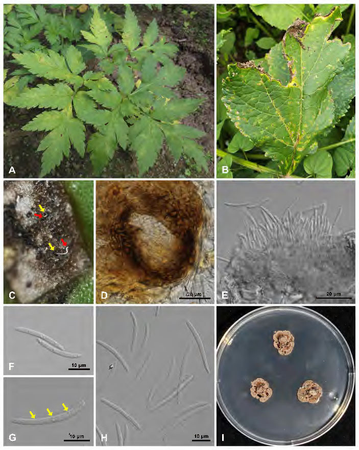
Fig. 1. Leaf spot symptoms caused by Septoria dearnessii on Heracleum moellendorffii. A, infected plants showing leaf spot symptoms in the field; B, diseased leaf; C, leaf lesion bearing pycnidial conidiomata (yellow arrows) and hyaline conidial tendrils (red arrows); D, ostiole of a pycnidial conidioma on the leaf surface; E, conidioma producing conidia; F, aseptate conidia; G, 3-septate conidia; H, conidia; I, colonies of S. dearnessii grown on potato dextrose agar (PDA) for 3 weeks.
Detailed morphological observations were conducted using a compound light microscope (Olympus BX53-32XDIC equipped with an Olympus DP74 camera; Olympus, Tokyo, Japan). Conidiomata formed on both leaf surfaces and developed subepidermally. They were dark brown, subglobose, and unilocular, measuring 50–110 ºm in diameter. The ostiole was circular, ruptured the epidermis at maturity, and measured 20–30 ºm in diameter (Fig. 1D). Conidia were cylindrical, straight to slightly curved, hyaline, and smooth-walled. They gradually tapered toward both ends, with a truncate base and rounded apex. Conidia were 0–3-septate, and measured 17–33 Septoria species infecting hosts within the Apiaceae.
For fungal isolation, conidia were collected from fresh leaf samples obtained from three locations and suspended in sterile distilled water. The suspension was spread onto 2% potato dextrose agar (PDA; Difco, Becton, Dickinson and Company, Franklin Lakes, NJ, USA) using a sterile loop. After incubation at 20℃ for 3 days, individual colonies were transferred to fresh PDA plates and incubated at 20℃. After 3 weeks of growth on PDA, colonies were pale orange with a wrinkled surface. The colony center was slightly raised, whereas the margins were irregular and lobate (Fig. 1I). Representative isolates were deposited in the Korea Agricultural Culture Collection (KACC) under accession numbers KACC 411252, KACC 411253, and KACC 411254.
Genomic DNA was extracted from fungal isolates cultured on PDA for 2 weeks at 20 ± 1℃ using the Quick-DNA Fungal/Bacterial Miniprep Kit (Zymo Research, Irvine, CA, USA). The internal transcribed spacer (ITS), large subunit (LSU) rDNA, translation elongation factor 1-alpha (tef1), and β-tubulin (Btub) regions were amplified using the primer pairs ITS5/ITS4 [11], LSU1Fd [12]/LR5 [13], EF1-728F [14]/EF-2 [15], and T1 [16]/β-Sandy-R [17], respectively. PCR amplification and reaction conditions followed previously published protocols [4]. The resulting amplicons were sequenced by a commercial sequencing service (Cosmogenetech, Seoul, Korea). Forward and reverse reads were examined and assembled using BioEdit version 7.0.5 [18] to obtain consensus sequences, which were subsequently deposited in GenBank. Table 1 presents the accession numbers.
To assess genetic similarity to known taxa, BLASTn searches were conducted for each gene region against the NCBI GenBank database. The ITS sequence shared 99.61% identity with S. dearnessii, while the LSU sequence exhibited high similarity to S. aegopodina (99.89%) and S. dearnessii (99.66%). In contrast, the Btub and tef1 regions showed relatively low sequence identities to reference strains of S. dearnessii, ranging from 96.23% to 97.06% and 93.39% to 95.27%, respectively.
The phylogenetic dataset included ITS, LSU, tef1, and β-tubulin sequences from 22 reference isolates, including two Cercospora species, retrieved from the NCBI GenBank database (Table 1). Cercospora apii and C. beticola were designated as outgroup taxa [5,9]. Each gene region was aligned individually using MAFFT and manually refined in BioEdit. The aligned sequences from the four loci were concatenated into a 1,930-character dataset. Maximum likelihood (ML) analysis was performed using RAxML v8.2.12 under the GTRCAT substitution model, with rapid bootstrap analysis and simultaneous search for the best-scoring ML tree. Branch support was evaluated using 1,000 bootstrap replicates, with values ≥70% considered to indicate significant support. The resulting phylogenetic tree was visualized and edited using MEGA 11 [19] (Fig. 2). In the multilocus phylogenetic analysis, the isolates formed a distinct lineage within the S. dearnessii clade, indicating close relatedness to S. dearnessii but a genetically differentiated lineage.
Table 1. List of fungal isolates used for phylogenetic analysis
| Species | Isolate no | Host | Location | GenBank accession number | |||
|---|---|---|---|---|---|---|---|
| ITS | LSU | tef1 | Btub | ||||
| Cercospora apii | CBS 118712 | – | Fiji | KF251296 | KF251800 | KF253244 | KF252778 |
| C. beticola | CBS 124.31 | Beta vulgaris | Romania | KF251146 | KF251650 | KF253106 | KF252645 |
| Septoria aegopodina | CBS 123740 | Aegopodium podagraria | Czech Republic | KF251335 | KF251839 | KF253282 | KF252807 |
| S. aegopodina | CBS 123741 | Aegopodium podagraria | Czech Republic | KF251334 | KF251838 | KF253281 | KF252806 |
| S. bupleuricola | CBS 128601 | Bupleurum lon giradiatum | South Korea | KF251355 | KF251859 | KF253302 | KF252827 |
| S. bupleuricola | CBS 128603 | Bupleurum falcatum | South Korea | KF251356 | KF251860 | KF253303 | KF252828 |
| S. campanulae | CBS 128589 | Campanula takesimana | South Korea | KF251360 | MH876464 | KF253307 | KF252832 |
| S. campanulae | CBS 128604 | Campanula takesimana | South Korea | KF251361 | KF251865 | KF253308 | KF252833 |
| S. coprosma | CBS 113391 | Coprosma robusta | New Zealand | KF251308 | KF251812 | KF253255 | KF252787 |
| S. dearnessii | KACC 411252 | Heracleum moellendorffii | South Korea | PX760817 | PX760825 | PZ121221 | PZ125585 |
| S. dearnessii | KACC 411253 | Heracleum moellendorffii | South Korea | PX760681 | PX760824 | PZ121222 | PZ125586 |
| S. dearnessii | KACC 411254 | Heracleum moellendorffii | South Korea | PX761787 | PX760826 | PZ125584 | PZ125587 |
| S. dearnessii | CBS 128624 | Angelica dahurica | South Korea | KF251400 | KF251904 | KF253347 | KF252871 |
| S. dearnessii | BCRC FU31532 (= R. Kirschner 4891) | Glehnia littoralis | Taiwan | MT843890 | – | LC574067 | LC574068 |
| S. gentianae | CBS 128633 | Gentiana scabra | South Korea | KF251426 | KF251930 | KF253374 | KF252898 |
| S. lactucae | CBS 108943 | Lactuca sativa | Netherlands | KF251439 | KF251943 | KF253387 | KF252911 |
| S. lactucae | CBS 352.58 | Lactuca sativa | Germany | KF251440 | KF251944 | KF253388 | KF252912 |
| S. mazi | CBS 128656 | Mazus japonicus | South Korea | KF251473 | KF251977 | KF253421 | KF252944 |
| S. mazi | CBS 128755 | Mazus japonicus | South Korea | KF251474 | KF251978 | KF253422 | KF252945 |
| S. oenanthes | CBS 128667 | Cicuta virosa | South Korea | KF251485 | KF251989 | KF253432 | KF252953 |
| S. oenanthicola | CBS 128649 | Oenanthe javanica | South Korea | KF251484 | KF251737 | KF253433 | KF252954 |
| S. sii | CBS 118.96 | Berula erecta | Netherlands | KF251550 | KF252055 | KF253498 | KF253018 |
| S. sii | CBS 102370 | Berula erecta | Netherlands | KF251549 | KF252054 | KF253497 | KF253017 |
| S. sonchi | CBS 128757 | Sonchus asper | South Korea | KF251552 | KF252057 | KF253500 | KF253020 |
| Septoria sp. | CBS 135474 | Conyza canadensis | Brazil | KF251559 | KF252064 | KF253507 | KF253027 |
ITS: internal transcribed spacer; LSU: large subunit ribosomal RNA; tef1: translation elongation factor 1-alpha; Btub: beta-tubulin; CBS: CBS Culture Collection, Westerdijk Fungal Biodiversity Institute, Utrecht, The Netherlands; KACC: Korean Agricultural Culture Collection, Rural Development Administration, Korea; BCRC: Bioresource Collection and Research Center, Taiwan;
Isolates obtained in this study and their newly generated sequences are indicated in bold.
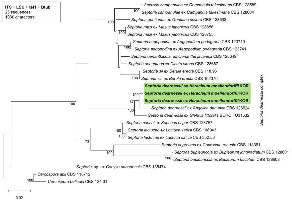
Fig. 2. Maximum likelihood phylogenetic tree of Septoria dearnessii inferred from a combined dataset of the internal transcribed spacer (ITS) region, the large subunit (LSU) rDNA, translation elongation factor 1-alpha (tef1), and β-tubulin (Btub) gene sequences. Bootstrap values ≥70% obtained from 1,000 replicates are shown at the nodes. The isolates obtained in this study are indicated in bold.
The genus Septoria exhibits host jumping, limiting the reliability of species delimitation based solely on host association [5]. The isolates obtained from H. moellendorffii showed morphological characteristics similar to those of S. dearnessii and several other Septoria species associated with Apiaceae hosts, including S. aegopodina, S. amphigena, S. bupleuricola, S. heracleicola, S. laubertiana, S. oenanthicola, S. oenanthis, S. pimpinellae, and S. sii [4,5,20–23]. However, multilocus phylogenetic analysis revealed that the isolates formed a lineage closely related to, but distinct from S. dearnessii (Fig. 2). Direct phylogenetic comparisons with other morphologically similar Septoria species remain limited owing to the lack of reference sequences. Furthermore, S. dearnessii represents a species complex rather than a single species [9]. Given their morphological similarity to S. dearnessii and the lack of molecular data for other morphologically similar Septoria species from Apiaceae hosts, the isolates are tentatively identified as S. dearnessii. Additional molecular data from S. dearnessii and related Apiaceae-associated Septoria species will be required to clarify their species-level taxonomic status. The morphological and molecular data presented in this study expand current knowledge of S. dearnessii and related Septoria species reported on Apiaceae hosts and provide a reference for future taxonomic clarification.
The authors declare that there are no conflicts of interest.
This work was supported by the National Institute of Forest Science under grant [Project No. FE0100-23-02-2026].
1. Youn WB, Hernandez JO, Park BB. Effects of shade and planting methods on the growth of Heracleum moellendorffii and Adenophora divaricata in different soil moisture and nutrient conditions. Plants 2021;10:2203. https://doi.org/10.3390/plants10102203
[DOI][PubMed][PMC]
2. Park HJ, Nugroho A, Jung B, Won YH, Jung YJ, Kim WB, Choi JS. Isolation and quantitative analysis of flavonoids with peroxynitrite-scavenging effect from the young leaves of Heracleum moellendorffii. Korean J Plant Res 2010;23:393-8.
3. Korea Forest Service. Production of forest products in 2024. Daejeon: Korea Forest Service; 2025.
4. Quaedvlieg W, Verkley GJM, Shin HD, Barreto RW, Alfenas AC, Swart WJ, Groenewald JZ, Crous PW. Sizing up Septoria. Stud Mycol 2013;75:307-90. https://doi.org/10.3114/sim0017
[DOI][PubMed][PMC]
5. Verkley GJM, Quaedvlieg W, Shin HD, Crous PW. A new approach to species delimitation in Septoria. Stud Mycol 2013;75:213-305. https://doi.org/10.3114/sim0018
[DOI][PubMed][PMC]
6. Ellis JB, Everhart BM. New and rare species of North American fungi (Sphaeropsidae). J Mycol 1889;5:145-57.
[DOI]
7. Sameva EF. New records of anamorphic fungi from Bulgaria. Mycol Balc 2004;1:55-7.
8. Shin HD, Sameva EF. Septoria in Korea. Suwon: National Institute of Agricultural Science and Technology; 2004. p. 183.
9. Yeh YH, Wu SH, Kirschner R. What is Septoria dearnessii? Plant Pathol Quar 2021;11:86-95. https://doi.org/10.5943/ppq/11/1/10
[DOI]
10. Cho WD, Shin HD. List of plant diseases in Korea. 4th ed. Seoul: Korean Society of Plant Pathology; 2004. p. 779.
11. White TJ, Bruns TD, Lee SB, Taylor JW. Amplification and direct sequencing of fungal ribosomal RNA genes for phylogenetics. In: Innis MA, Gelfand DH, Sninsky JJ, White TJ, editors. PCR protocols: A guide to methods and applications. San Diego: Academic Press; 1990. p. 315-22. https://doi.org/10.1016/B978-0-12-372180-8.50042-1
[DOI][PubMed][PMC]
12. Crous PW, Schoch CL, Hyde KD, Wood AR, Gueidan C, de Hoog GS, Groenewald JZ. Phylogenetic lineages in the Capnodiales. Stud Mycol 2009;64:17-47. https://doi.org/10.3114/sim.2009.64.02
[DOI][PubMed][PMC]
13. Vilgalys R, Hester M. Rapid genetic identification and mapping of enzymatically amplified ribosomal DNA from several Cryptococcus species. J Bacteriol 1990;172:4238-46. https://doi.org/10.1128/jb.172.8.4238-4246.1990
[DOI][PubMed][PMC]
14. Carbone I, Kohn LM. A method for designing primer sets for speciation studies in filamentous ascomycetes. Mycologia 1999;91:553-6. https://doi.org/10.1080/00275514.1999.12061051
[DOI]
15. O’Donnell K, Kistler HC, Cigelnik E, Ploetz RC. Multiple evolutionary origins of the fungus causing Panama disease of banana: Concordant evidence from nuclear and mitochondrial gene genealogies. Proc Natl Acad Sci USA 1998;95:2044-9. https://doi.org/10.1073/pnas.95.5.2044
[DOI][PubMed][PMC]
16. O’Donnell K, Cigelnik E. Two divergent intragenomic rDNA ITS2 types within a monophyletic lineage of the fungus Fusarium are nonorthologous. Mol Phylogenet Evol 1997;7:103-16. https://doi.org/10.1006/mpev.1996.0376
[DOI][PubMed]
17. Stukenbrock EH, Quaedvlieg W, Javan-Nikhah M, Zala M, Crous PW, McDonald BA. Zymoseptoria ardabiliae and Z. pseudotritici, two progenitor species of the Septoria tritici leaf blotch fungus Z. tritici (synonym: Mycosphaerella graminicola). Mycologia 2012;104:1397-407. https://doi.org/10.3852/11-374
[DOI][PubMed]
18. Hall TA. BioEdit: A user-friendly biological sequence alignment editor and analysis program for Windows 95/98/NT. Nucleic Acids Symp Ser 1999;41:95-8.
19. Tamura K, Stecher G, Kumar S. MEGA11: Molecular evolutionary genetics analysis version 11. Mol Biol Evol 2021;38:3022-7. https://doi.org/10.1093/molbev/msab120
[DOI][PubMed][PMC]
20. Fedyushko IA. Key to phytopathogenic pycnidial Septoria-like micromycetes of Belarus. Minsk: Belarusian State University; 2025. p. 525.
21. Radulescu E, Negru A, Docea E. Septoriozele din Romania. Bucuresti: Editura Academiei Republicii Socialiste Romania; 1973.
22. Saccardo PA, Saccardo D, Traverso JB, Trotter A. Sylloge Fungorum. Vol. 25. Padova: Sumptibus Auctorum; 1931. p. 456.
23. Miyake I. Studies in Chinese fungi (with plate I). Bot Mag (Tokyo) 1912;26:51-66.
[DOI]