
Ju-Heon Lee1,2, Youngsoo Kim1, Jong-Taek Park1, Dong-Hyuk Lee1, Hye-Yeon Mun3, Seung-Yoon Oh4, and Hee-Young Jung2*
1Apple Research Center, National Institute of Horticultural & Herbal Science, Gunwi 43100, Korea
2Department of Plant Medicine, Kyungpook National University, Daegu 41566, Korea
3Fungal Research Team, Nakdonggang National Institute of Biological Resources, Sangju 37242, Korea
4Department of AI-Integrated Biological Sciences, Changwon National University, 20 Changwondaehak-ro, Changwon 51140, Korea
*Correspondence to heeyoung@knu.ac.kr
Korean Journal of Mycology (Kor J Mycol) 2026 June, Volume 54, Issue 2, pages 207-218.
https://doi.org/10.4489/kjm.2026.54.2.10
Received on June 09, 2026, Revised on June 26, 2026, Accepted on June 30, 2026, Published on June 30, 2026.
Copyright © The Korean Society of Mycology.
This is an Open Access article which is freely available under the Creative Commons Attribution-Non-Commercial 4.0 International License (CC BY-NC) (https://creativecommons.org/licenses/by-nc/4.0/).
Ambrosia beetles, Coniochaeta luteoviridis, Morphology, Phylogeny
The genus Coniochaeta comprises a pleomorphic group of fungi within the family Coniochaetaceae [1], with Coniochaeta ligniaria designated as the type species. Currently, more than 100 species are recognized within the genus [2,3]. In earlier taxonomic frameworks, Coniochaeta was regarded as the sexual morph, whereas Lecythophora represented the corresponding asexual morph and was treated as a separate genus. Following the abolition of the dual nomenclature system for pleomorphic fungi, the older name Coniochaeta was conserved under the principle of nomenclatural priority, and species formerly assigned to Lecythophora were transferred to Coniochaeta [4].
Morphologically, Coniochaeta species form white to salmon-colored or yellowish colonies, which may darken to olive or brown in the later stages of cultivation. Their conidia are produced phialidically and are ellipsoidal to cylindrical, and some species form globose to ellipsoidal chlamydospores [1].
Species of Coniochaeta have been documented across diverse ecological habitats, including wood, freshwater environments, and plant tissues, and have even been isolated from extreme environments such as uranium mines [3–5]. Numerous studies indicate that many members of the genus form endophytic associations with plants and typically remain asymptomatic. For instance, Coniochaeta spp. have been isolated from Panax notoginseng [6], Coniochaeta endophytica has been reported from healthy photosynthetic tissues of Platycladus orientalis, and Coniochaeta ligniaria has been isolated from leaves of Baeckea frutescens. Taken together, these findings indicate that Coniochaeta species predominantly exhibit an endophytic lifestyle [2,7].
Despite their frequent occurrence as endophytes, certain Coniochaeta species have been implicated in human infections. Notably, C. hoffmannii and C. mutabilis are recognized as opportunistic human pathogens and have been implicated in keratitis, subcutaneous abscesses, peritonitis, and endocarditis [8]. Moreover, several newly described Coniochaeta species originating from human and veterinary sources have been reported in recent years [9,10].
In Korea, the first case of fungemia caused by C. hoffmannii has been documented [11]. Beyond clinical occurrences, C. ligniaria has been isolated from leaves of Abies koreana [12], and C. velutina has been detected in indoor air samples, indicating a gradual expansion of domestic research on this genus.
In the present study, a Coniochaeta-like fungus was isolated from an ambrosia beetle known to cause damage to apple trees. To clarify its taxonomic placement, we conducted detailed morphological and molecular phylogenetic analyses, enabling species-level identification.
Ambrosia beetles were collected using traps installed in an orchard at the Apple Research Center, Gunwi-gun, Daegu-si, Republic of Korea (36°16′44.1″N, 128°27′56.2″E). Collected individuals were surface-sterilized by immersion in 70% ethanol for 1 min and subsequently air‑dried at room temperature for approximately 10 min. Following surface sterilization, individual beetles were placed onto potato dextrose agar (PDA; Difco, Detroit, MI, USA) plates and incubated at 25°C. After 3 days of incubation, emerging fungal mycelia were transferred to fresh PDA plates and incubated for an additional 10 days at the same temperature to obtain pure cultures. The resulting single isolate was designated ARI-25-A13 and preserved in 20% glycerol at −80°C for long-term storage. In addition, the isolate was deposited in the Korean Agricultural Culture Collection (KACC) under accession number KACC 411349.
Isolate ARI-25-A13 was cultured on PDA and malt extract agar (MEA; Difco, Detroit, MI, USA) media at 25°C for 14 days to assess its cultural and morphological characteristics. Colony diameter, pigmentation, and developmental changes were recorded throughout incubation. In addition, the morphological features of conidia and conidiophores were observed using a light microscope (CX-43, Olympus, Tokyo, Japan), and the dimensions of each structure were measured.
Genomic DNA was extracted from isolate ARI-25-A13 using the HiGene genomic DNA preparation kit (Biofact, Daejeon, Korea) following the manufacturer’s protocol. Subsequently, the internal transcribed spacer (ITS) region and the large subunit of nuclear ribosomal RNA (LSU) region were sequenced to infer the genus-level identity of ARI-25-A13. For species-level identification, partial sequences of the translation elongation factor 1-alpha (TEF1-α), β-tubulin (TUB), and actin (ACT) genes were amplified by polymerase chain reaction (PCR).
The ITS region was amplified using primers ITS1F/ITS4 [13,14], and the LSU gene using LR0R/LR7 [15]. The TEF1-α gene was amplified with primers 983f/2218R [16], whereas TUB and ACT were amplified using BT1819R/BT2916 [17] and ACT-2/ACT-4r [18], respectively.
PCR products were separated on 1% agarose gels and visualized by ethidium bromide staining. Amplified fragments were purified using EXOSAP-IT reagent (Thermo Fisher Scientific, Waltham, MA, USA) in accordance with the manufacturer’s protocol, and sequenced by Solgent Co., Ltd. (Daejeon, Korea).
Sequence assembly and analysis were carried out using SeqMan Lasergene software (DNAStar Inc., Madison, WI, USA). The ITS (LC928497), LSU (LC928498), TEF1-α (LC928499), TUB (LC928501), and ACT (LC928500) sequences obtained in this study were deposited in GenBank, with their accession numbers listed in Table 1.
Table 1. List of Coniochaeta species included in the phylogenetic analyses and their GenBank accession numbers
| Species | Isolate | Isolation source | GenBank accession numbers | |||
|---|---|---|---|---|---|---|
| ITS | LSU | TUB | ACT | |||
| Coniochaeta acaciae | MFLUCC 17-2298T | Dead branches of Fabaceae | MG062735 | MG062737 | – | – |
| Coniochaeta arenariae | MFLUCC 18-0409 | Ammophila arenaria | MN047126 | MN017896 | – | – |
| Coniochaeta africana | CBS 120868T | Prunus salicina | NR_137725 | NG_066150 | – | – |
| Coniochaeta baysunika | MFLUCC 17-0830T | Rosa sp. | MG828880 | MG828996 | – | – |
| Coniochaeta boothii | CBS 381.74T | Soil | NR_159776 | AJ875226 | – | – |
| Coniochaeta cateniformis | UTHSC 01-1644T | Canine bone marrow | NR_111517 | HE610329 | HE610347 | HE610339 |
| Coniochaeta canina | UTHSC 11-2460T | Canine breed German Shepard | NR_120211 | NG_042720 | – | – |
| Coniochaeta coluteae | MFLUCC 17-2299T | Colutea paulsenii | MG137251 | MG137252 | – | – |
| Coniochaeta decumbens | CBS 153.42T | Fruit | NR_144912 | AF353597 | – | – |
Coniochaeta deborreae | CBS 147215T | Soil | NR_173009 | NG_076709 | – | – |
| Coniochaeta discoidea | CBS 158.80T | Soil | NR_159779 | NG_064120 | – | – |
| Coniochaeta endophytica | AEA 9094T | Platycladus orientalis | EF420005 | NG_075158 | – | – |
| Coniochaeta euphorbiae | CBS 139768T | Euphorbia polycaulis | KP941076 | KP941075 | – | – |
| Coniochaeta fasciculata | CBS 205.38T | Butter | NR_154770 | AF353598 | HE610350 | HE610342 |
| Coniochaeta fibrosae | CGMCC3.20304T | Candelaria fibrosa | MW750760 | MW750758 | – | – |
| Coniochaeta fodinicola | CBS 136963T | Uranium mine | JQ904603 | KF857172 | – | – |
| Coniochaeta gigantospora | ILLS 60816T | Fraxinus Excelsior wood submerged In the river | NR_121521 | JN684909 | – | – |
| Coniochaeta hoffmannii | CBS 245.38T | Butter | NR_167688 | MH867452 | – | – |
| Coniochaeta iranica | CBS 139767T | Euphorbia polycaulis | KP941078 | KP941077 | – | – |
| Coniochaeta ligniaria | CBS 424.65T | Picea abies | MH858650 | MH870292 | – | – |
| Coniochaeta lignicola | CBS 267.33T | Unknown | NR_111520 | NG_067344 | HE610353 | HE610345 |
| Coniochaeta lignicola | CBS 590.63 | Decaying wood | OQ429536 | OQ055448 | ||
| Coniochaeta luteorubra | CBS 131710T | Leg wound | HE610330 | HE610328 | HE610346 | HE610338 |
| Coniochaeta luteoviridis | CBS 206.38T | Picea abies | NR_154769 | NG_067348 | HE610351 | HE610343 |
| Coniochaeta luteoviridis | IFM 50534 | Unknown | AB190401 | AB190429 | ||
| Coniochaeta luteoviridis* | ARI-25-A13 | Ambrosia beetle | LC928497 | LC928498 | LC928501 | LC928500 |
| Coniochaeta marina | MFLUCC 18-0408T | From the sea, Leif Tibell | MK458764 | MK458765 | – | – |
| Coniochaeta monsterae | URM 8284T | Leaf endophytic Monstera dansonii | MZ648895 | MZ648891 | – | – |
| Coniochaeta mongoliae | CGMCC3.20250T | Medulla of Ramalina sinensis | MW077645 | MW077646 | – | – |
| Coniochaeta mutabilis | CBS 157.44T | Picea abies | NR_111519 | NG_042382 | HE610349 | HE610341 |
| Coniochaeta navarrae | CBS 141016T | On bark of Ulmus sp. | NR_154808 | KU762326 | – | – |
| Coniochaeta ostrea | CBS 507.70T | Twig of Larrea sp. | NR_159772 | NG_064080 | – | – |
| Coniochaeta polymorpha | CBS 132722T | Endotracheal aspirate of preterm Neonate | NR_121473 | HE863327 | HE863708 | HE863709 |
| Coniochaeta polysperma | CBS 669.77T | Dung of hare | MH861109 | MH872868 | – | – |
| Coniochaeta prunicola | CBS 120875T | Prunus armeniaca | NR_137037 | NG_066151 | – | – |
| Coniochaeta prunicola | HMJAU 34702 | Rotten wood | MZ346578 | MZ346514 | – | – |
| Coniochaeta rosae | TASM 6127T | Rosa hissarica | NR_157509 | NG_066204 | – | – |
| Coniochaeta rosae | MFLUCC 17-0806 | Branches | MG828882 | MG828998 | ||
| Coniochaeta rhopalochaeta | CBS 109872T | Bulnesia retamas | NR_172554 | GQ351561 | – | – |
| Coniochaeta savoryi | CBS 725.74T | Wood of Juniperus scopulorum | MH860890 | MH872627 | – | – |
| Coniochaeta simbalensis | NFCCI 4236T | Soil | NR_164024 | NG_068555 | – | – |
| Coniochaeta sinensis | CGMCC3.20306T | Medulla of Ramalina sinensis | MW422269 | MW422265 | – | – |
| Coniochaeta velutina | CBS 981.68 | bark beetle galleries of Hylurgops palliatus | MH859264 | MH870991 | – | – |
| Coniochaeta velutina | HMJAU 34693 | Rotten wood | MZ346570 | MZ346506 | – | – |
| Coniochaeta vineae | KUMCC 17-0322T | On dead vine | NR_168225 | MN473512 | – | – |
| Coniochaeta verticillata | CBS 816.71T | Soil | NR_159774 | AJ875232 | – | – |
| Phialemonium obovatum | CBS 279.76T | Man, systemic mycotic infection in a six-month-old burned child | NR_165935 | NG_057631 | LT634002 | HE599315 |
ITS, internal transcribed spacer; LSU, large subunit ribosomal DNA; TUB, β-tubulin; ACT, actin.
T: ex-type.
*The strain isolated in this study is shown in bold.
Each taxonomic marker sequence was subjected to a Basic Local Alignment Search Tool (BLAST) search against the NCBI database to assess sequence similarity. Based on these results, closely related strains were selected, and their sequence homology with ARI‑25‑A13 was evaluated.
The BLAST-based homology results were used to infer the genus to which ARI-25-A13 belongs and to guide the selection of molecular markers for phylogenetic reconstruction. Sequence alignment and phylogenetic analyses were conducted in MEGA 7 [19]. The sequences of the selected molecular markers were aligned using Clustal W, and the aligned sequences of the ITS, LSU, TUB, and ACT genes were concatenated for multilocus phylogenetic analysis.
Phylogenetic analysis was performed using the maximum likelihood (ML) method [20] under the Kimura two-parameter model [21], with gap positions excluded from the analysis. The reliability of the inferred phylogenetic tree was assessed by bootstrap analysis with 1,000 replicates.
On PDA at 25°C, colonies of ARI‑25‑A13 reached 20.3–21.2 mm in diameter after 7 days and 36.7–37.4 mm after 14 days. Colonies initially appeared salmon-colored and gradually transitioned to olive to brown as incubation progressed (Fig. 1A). On MEA under identical conditions, colony diameters were 9.0–9.5 mm after 7 days and 16.3–17.3 mm after 14 days. Colonies retained a salmon coloration throughout the incubation period (Fig. 1B). Adelophialides were short, thin-walled, and inconspicuous. Collarettes were not observed, and discrete phialides were absent (Fig. 1C–E). Conidia were produced in small groups of 2–4 at the apex. Chlamydospores were hyaline, globose to ellipsoidal, and formed terminally or intercalarily, occurring singly or in short chains. They measured 4.0–8.0 × 3.3–4.5 μm (mean 5.8 × 4.0 μm, Q = 1.5, n = 10) (Fig. 1F–G). Conidia were hyaline, ellipsoidal to cylindrical, frequently slightly curved, and measured 3.7–5.9 × 1.4–2.6 μm (mean 4.7 × 1.9 μm, mean Q = 2.5, n = 100) (Fig. 1H). Yellow-pigmented hyphae were occasionally observed and appeared as aggregated hyphal masses (Fig. 1I).
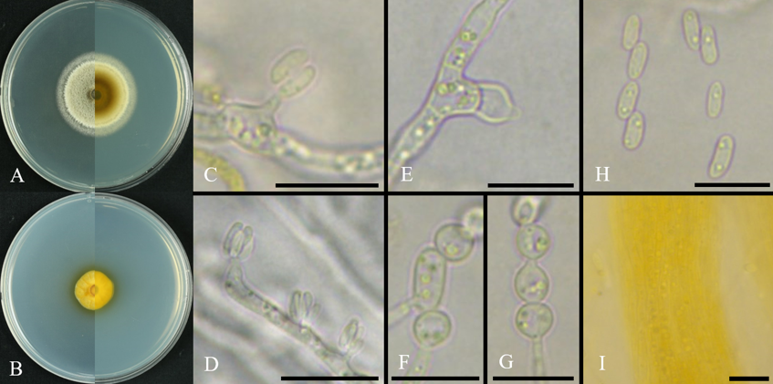
Fig. 1. Cultural and morphological characteristics of Coniochaeta luteoviridis ARI-25-A13. A: Front and reverse views of the colony grown on potato dextrose agar (PDA) for 14 days at 25°C. B: Front and reverse views of the colony grown on malt extract agar (MEA) for 14 days at 25°C. C–E: Adelophialides. F–G: Chlamydospores. H: Conidia. I: Yellow-pigmented hyphal aggregates. Scale bars: C–I = 10 μm.
To elucidate the molecular and phylogenetic relationships of ARI-25-A13, sequences of the ITS and LSU regions and of the TEF1-α, ACT, and TUB genes were analyzed. Each sequence was compared with reference strains deposited in the NCBI database using BLAST searches.
The ITS sequence (586 bp) showed 100.0% similarity to Coniochaeta luteoviridis KoRLI047395 (GenBank no. MN341269), followed by C. rosae 4.27A (OP584633; 99.7%), C. weberae JKI-GP-23-051 (PV272675; 99.7%), C. luteoviridis CBS 206.38T (MH855948; 99.3%), and C. fasciculata GSVCC22R3-2 (OR400651; 98.6%). The LSU sequence (1,289 bp) showed 100% similarity to Coniochaeta luteoviridis CBS 206.38T (NG_067348.1), followed by C. velutina UAMH 10912 (EU999180; 99.8%), C. cephalothecoides L821 (KY064030; 99.1%), C. hoffmannii CBS 245.38T (MG491499; 99.0%), and C. endophytica AEA 9094T (NG_075158; 99.0%).
The TEF1-α sequence (905 bp) showed 98.8% similarity to Coniochaeta lignicola CBS 590.63 (OQ470829), followed by C. fasciculata CBS 205.38T (MK693152; 98.7%) and C. velutina HMJAU 34696 (MZ465111; 98.5%). The ACT sequence (647 bp) showed the highest similarity to Coniochaeta luteoviridis CBS 206.38T (HE610343; 99.0%), followed by C. velutina HMJAU 34693 (MZ464934; 96.2%), C. lignicola CBS 267.33T (HE610345; 95.3%), and C. fasciculata CBS 205.38T (HE610342; 95.3%). The TUB sequence (1,159 bp) showed the highest similarity to Coniochaeta fasciculata CBS 205.38T (HE610350; 96.5%), followed by C. luteoviridis CBS 206.38T (HE610351; 96.2%) and C. lignicola CBS 267.33T (HE610353; 94.7%).
To determine the phylogenetic placement of ARI-25-A13, an ML phylogeny was constructed using concatenated ITS and LSU sequences. ARI-25-A13 clustered most closely with C. luteoviridis CBS 206.38T with 76% bootstrap support, whereas its separation from the closely related C. lignicola was not strongly resolved in the combined ITS and LSU phylogeny (Fig. 2). To further refine its taxonomic position, a multilocus phylogenetic analysis was conducted using ITS, LSU, TUB, and ACT sequences. The resulting ML phylogeny showed that ARI‑25‑A13 placed in a close relationship with C. luteoviridis CBS 206.38T and was clearly separated from other species, including C. lignicola (Fig. 3).
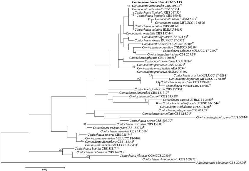
Fig. 2. Combined internal transcribed spacer (ITS) and large subunit ribosomal DNA (LSU) sequence data were used to infer a maximum-likelihood phylogenetic tree of Coniochaeta, in which the position of ARI-25-A13 is shown. Bootstrap values exceeding 70% from 1,000 replications are provided at the nodes. The isolate obtained in this study is indicated in bold. Phialemonium obovatum CBS 279.76T was included as the outgroup. Scale bar = 0.02 substitutions per nucleotide position. ‘T’: ex-type.
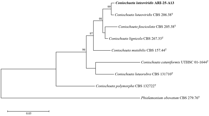
Fig. 3. Maximum-likelihood phylogenetic tree inferred from the combined internal transcribed spacer (ITS), large subunit ribosomal DNA (LSU), β-tubulin (TUB), and actin (ACT) sequences, showing the phylogenetic position of ARI-25-A13 among species of the genus Coniochaeta. Bootstrap values above 70% from 1,000 replications are presented at the nodes. The isolate obtained in this study is shown in bold. Phialemonium obovatum CBS 279.76T was used as the outgroup. The scale bar indicates 0.05 substitutions per nucleotide position. ‘T’: ex-type.
Although ARI-25-A13 was phylogenetically related to both Coniochaeta luteoviridis and C. lignicola, its morphological characteristics more closely matched those of C. luteoviridis, whereas it was clearly distinguishable from C. lignicola.
On PDA, ARI‑25‑A13 exhibited an initial salmon coloration that gradually shifted to olive to brown with age, consistent with the documented color transition of C. luteoviridis CBS 206.38T from salmon to olivaceous to brown on MEA (Table 2). In addition, ARI-25-A13 showed high similarity to C. luteoviridis CBS 206.38T in conidial morphology, adelophialide characteristics, the absence of discrete phialides, and the presence and mode of formation of chlamydospores. Conidia of ARI-25-A13 (3.7–5.9 × 1.4–2.6 μm) were similar in shape to those of C. luteoviridis CBS 206.38T (4.5–7 × 1.8–2.5 μm), although they were slightly smaller.
In contrast, C. lignicola has been reported to produce colonies that darken progressively with age, a pattern distinct from that of ARI-25-A13. Its conidia are described as ovoid to ellipsoidal, which distinguishes them from the somewhat curved conidia observed in ARI-25-A13. Conidia of C. lignicola CBS 267.33T (3.0–4.5 × 1.5–2.0 μm) are also smaller and more uniform in shape than those of ARI-25-A13. Moreover, C. lignicola CBS 267.33T is clearly distinguished from ARI-25-A13 by its conidiophore characteristics, particularly the predominance of discrete ventricose phialides and the presence of relatively long collarettes. The absence of chlamydospores in C. lignicola represents an additional diagnostic feature separating it from ARI-25-A13 (Table 2).
Table 2. Comparison of the morphological characteristics of isolate ARI-25-A13 with previously reported descriptions of Coniochaeta luteoviridis CBS 206.38T and C. lignicola CBS 267.33T
| Characteristics | C. luteoviridis ARI-25-A13a | C. luteoviridis CBS 206.38Tb | C. lignicola CBS 267.33Tb | |
|---|---|---|---|---|
| Colony | Color, Shape | Colony on PDA: initially salmon-colored, becoming olive to brown with age. Colony on MEA: remaining salmon-colored throughout the incubation period. | Colony on MEA: Initially salmon, later olivaceous to brown, surface covered with abundant aerial mycelium. | Colonies darkening with age. |
Size (mm) | Colonies on PDA attaining 36.7–37.4 mm diam after 14 days at 25°C; colonies on MEA attaining 16.3–17.3 mm diam after 14 days at 25°C. | Colonies on MEA attaining 29.0 mm diam after 14 days. | Colonies on MEA attaining 30.0–35.0 mm diam after 14 days. | |
| Conidiophores | Color | Hyaline | N/A | N/A |
| Shape | Adelophialides short, thin-walled, and inconspicuous, collarettes not observed, discrete phialides absent. | Adelophialides thin-walled and inconspicuous, with inconspicuous collarettes; discrete phialides not observed. | Adelophialides present but less abundant than discrete ventricose phialides, which are dominant and bear relatively long collarettes (1–1.5(–2) μm), cylindrical to slightly widened. | |
| Conidia | Color | Hyaline | N/A | N/A |
| Shape | Ellipsoidal to cylindrical, often slightly curved. | Ellipsoidal to cylindrical, sometimes slightly curved, containing small guttules. | Ovoidal to ellipsoidal, relatively small and regular in shape. | |
Size (μm) | 3.7–5.9 × 1.4–2.6 | 4.5–7.0 × 1.8–2.5 | 3.0–4.5 × 1.5–2.0 | |
| Chlamydospores | Color | Hyaline | Hyaline to faintly brown. | Not observed. |
| Shape | Globose to ellipsoidal, formed terminally or intercalarily, occurring singly or in short chains. | Globose to ellipsoidal or pear-shaped, slightly thick-walled, terminal or intercalary, occurring singly or in short chains. | ||
Size (μm) | 4.0–8.0 × 3.3–4.5 | 4.0–13.0 × 3.5–6.0 |
Tex-type strain; aFungal strain studied in this paper; bSource of description [1].
In this study, ambrosia beetles were collected using traps deployed in an apple orchard, and strain ARI-25-A13 was isolated from these insects. Sequence analysis of the ITS rDNA region indicated that the isolate belonged to the genus Coniochaeta, and an integrated assessment of its morphological traits and multilocus phylogenetic relationships ultimately supported its identification as Coniochaeta luteoviridis.
Currently, approximately 170 species of Coniochaeta are registered in the MycoBank database. Despite the broad ecological distribution of this genus, species‑level resolution based solely on ITS sequences remains challenging, as closely related taxa often cannot be reliably distinguished using this marker alone. Therefore, additional loci are required to achieve accurate species delimitation [2].
Recent studies on the classification and identification of novel Coniochaeta species have employed various molecular markers. Silva et al. [22] characterized Coniochaeta monsterae using ITS and LSU sequences, whereas Arnold et al. [23] used ITS and TEF1-α sequences to describe the new species Coniochaeta elegans, C. montana, and C. nivea, highlighting the utility of these loci for species‑level identification within Coniochaeta. In addition, Kabtani et al. [3] employed ITS, LSU, TUB, and TEF1-α sequences for the classification and description of C. massiliensis as a novel species. Accordingly, in the present study, ITS, LSU, TUB, and TEF1-α, which have been frequently used in recent identification studies, were initially selected for the molecular characterization of ARI-25-A13.
Phylogenetic analyses based on the ITS and LSU genes revealed that ARI-25-A13 was most closely related to C. luteoviridis, although the phylogeny did not clearly resolve its separation from the closely related C. lignicola. To refine this placement, additional loci were considered; however, because the TEF1-α sequence of the type strain of C. luteoviridis was not available in the NCBI database, this marker could not be incorporated in the final phylogenetic analysis. As an alternative, the ACT and TUB genes, which were used by Perdomo et al. [9] for the description and identification of the novel species C. luteorubra and C. cateniformis, were selected and included in the multilocus phylogenetic analysis. However, ACT and TUB sequences are not available for many Coniochaeta species in the NCBI database, which limited the number of comparable strains. Nevertheless, all available strains possessing ACT and TUB sequence data were included in the analysis, allowing the phylogenetic position of ARI-25-A13 to be evaluated in relation to C. luteoviridis and C. lignicola. Ultimately, the multilocus phylogenetic analysis based on ITS, LSU, TUB, and ACT sequences showed that ARI-25-A13 consistently clustered with C. luteoviridis with strong statistical support.
Although ARI-25-A13 showed phylogenetic affinity to both C. luteoviridis and C. lignicola, its morphological characteristics were overall more congruent with those of C. luteoviridis. In particular, the pattern of colony color change on PDA, conidial morphology, adelophialide structure, the absence of discrete phialides, and the presence and mode of formation of chlamydospores all closely matched the diagnostic features of C. luteoviridis.
However, a difference between ARI-25-A13 and C. luteoviridis CBS 206.38T was observed in growth rate on MEA. In the present study, ARI-25-A13 showed a colony diameter of 16.3–17.3 mm on MEA after 14 days, whereas that of C. luteoviridis CBS 206.38T was reported as 29.0 mm, indicating a notable difference in growth rate between the two strains. Although cultural characteristics have been considered in previous studies for distinguishing species of Coniochaeta, diagnostic traits such as colony color change, the morphology of conidiogenous cells, conidial morphology, and the presence or absence of chlamydospores have been regarded as more important than growth rate alone [4,22,24,25]. In addition, Weber [1] distinguished C. luteoviridis CBS 206.38T from C. velutina mainly based on the presence of chlamydospores in C. luteoviridis, despite their overall morphological similarities. Therefore, the difference in growth rate on MEA observed in this study may have been influenced by differences in medium composition. Considering the overall agreement in morphological characteristics and multilocus phylogenetic results, ARI-25-A13 is best assigned to C. luteoviridis.
In Korea, a previous study investigated the distribution and diversity of endolichenic fungi from Jeju Island based on ITS sequence data and reported the occurrence of five Coniochaeta species, including C. luteoviridis [26]. However, species-level identification among closely related taxa may be limited when relying solely on the ITS region. In contrast, the present study clarified the phylogenetic position of ARI-25-A13 more precisely through multilocus phylogenetic analysis using ITS, LSU, TUB, and ACT sequences, and further presents the morphological characteristics of C. luteoviridis.
ARI-25-A13 was isolated from an ambrosia beetle. In general, ambrosia beetles are known to form symbiotic associations with fungi such as Raffaelea spp. and Ambrosiella spp. These insects typically colonize woody hosts, inoculate them with their symbiotic fungi, and subsequently utilize the fungal growth as a primary nutritional resource [27]. Species of Coniochaeta have also been isolated from wind-thrown stems of Picea abies, including Coniochaeta ligniaria, C. velutina, and C. pulveracea, and in particular, C. malacotricha has been reported from bark beetles and their galleries associated with Picea and Pinus [1]. However, current evidence is insufficient to determine whether species of Coniochaeta form a symbiotic relationship with ambrosia beetles or were merely isolated incidentally as endophytic fungi. Accordingly, it would be premature to exclude the possibility of an ecological association between these fungi and their insect hosts. Further studies and continued isolation and identification efforts will be necessary to clarify their ecological association.
The authors declare no conflict of interest.
This work was supported by the “Cooperative Research Program for Agriculture Science and Technology Development (Project No. PJ017183)” funded by the Rural Development Administration, Republic of Korea. Additional support was provided by a grant from the Nakdonggang National Institute of Biological Resources (NNIBR), funded by the Ministry of Environment (MOE) of the Republic of Korea (NNIBR20261103).
1. Weber E. The Lecythophora-Coniochaeta complex: I. Morphological studies on Lecythophora species isolated from Picea abies. Nova Hedwig 2002;74:159-85. https://doi.org/10.1127/0029-5035/2002/0074-0159
[DOI]
2. Harrington AH, del Olmo-Ruiz M, U’Ren JM, Garcia K, Pignatta D, Wespe N, Sandberg DC, Huang YL, Hoffman MT, Arnold AE. Coniochaeta endophytica sp. nov., a foliar endophyte associated with healthy photosynthetic tissue of Platycladus orientalis (Cupressaceae). Plant Fungal Syst 2019;64:65-79. https://doi.org/10.2478/pfs-2019-0008
[DOI]
3. Kabtani J, Militello M, Ranque S. Coniochaeta massiliensis sp. nov. isolated from a clinical sampl28. J Fungi 2022;8:999. https://doi.org/10.3390/jof8100999
[DOI][PubMed][PMC]
4. Khan Z, Gené J, Ahmad S, Cano J, Al-Sweih N, Joseph L, Chandy R, Guarro J. Coniochaeta polymorpha, a new species from endotracheal aspirate of a preterm neonate, and transfer of Lecythophora species to Coniochaeta. Antonie van Leeuwenhoek 2013;104:243-52. https://doi.org/10.1007/s10482-013-9943-z
[DOI][PubMed]
5. Vázquez-Campos X, Kinsela AS, Waite TD, Collins RN, Neilan BA. Fodinomyces uranophilus gen. nov. sp. nov. and Coniochaeta fodinicola sp. nov., two uranium mine-inhabiting Ascomycota fungi from northern Australia. Mycologia 2014;106:1073-89. https://doi.org/10.3852/14-013
[DOI][PubMed]
6. Wei G, Chen Z, Wang B, Wei F, Zhang G, Wang Y, Zhu G, Zhou Y, Zhao Q, He M, et al. Endophytes isolated from Panax notoginseng converted ginsenosides. Microb Biotechnol 2021;14:1730-46. https://doi.org/10.1111/1751-7915.13842
[DOI][PubMed][PMC]
7. Kokaew J, Manoch L, Worapong J, Chamswarng C, Singburaudom N, Visarathanonth N, Piasai O, Strobel G. Coniochaeta ligniaria an endophytic fungus from Baeckea frutescens and its antagonistic effects against plant pathogenic fungi. Thai J Agric Sci 2011;44:123-31.
8. de Hoog GS, Guarro J, Gené J, Figueras MJ. Atlas of clinical fungi. 2nd ed. Utrecht: Centraalbureau voor Schimmelcultures; 2000.
9. Perdomo H, García D, Gené J, Cano J, Sutton DA, Summerbell R, Guarro J. Phialemoniopsis, a new genus of Sordariomycetes, and new species of Phialemonium and Lecythophora. Mycologia 2013;105:398-421. https://doi.org/10.3852/12-137
[DOI][PubMed]
10. Troy GC, Panciera DL, Pickett JP, Sutton DA, Gene J, Cano JF, Guarro J, Thompson EH, Wickes BL. Mixed infection caused by Lecythophora canina sp. nov. and Plectosphaerella cucumerina in a German shepherd dog. Med Mycol 2013;51:455-60. https://doi.org/10.3109/13693786.2012.754998
[DOI][PubMed]
11. Hong Y, Kim D, Park K, Sung H, Kim MN. The first case of Coniochaeta hoffmannii fungemia and literature review. Lab Med Online 2022;12:195-200. https://doi.org/10.47429/lmo.2022.12.3.195
[DOI]
12. Park H, Kim DY, Eom AH. Two unreported species of endophytic fungi isolated from leaves of Abies koreana in Korea. Kor J Mycol 2018;46:22-7. https://doi.org/10.4489/KJM.20180003
[DOI]
13. Gardes M, Bruns TD. ITS primers with enhanced specificity for basidiomycetes-application to the identification of mycorrhizae and rusts. Mol Ecol 1993;2:113-8. https://doi.org/10.1111/j.1365-294x.1993.tb00005.x
[DOI][PubMed]
14. White TJ, Bruns TD, Lee SB, Taylor JW. Amplification and direct sequencing of fungal ribosomal RNA genes for phylogenetics. In: Innis MA, Gelfand DH, Sninsky JJ, White TJ, editors. PCR protocols: A guide to methods and applications. San Diego: Academic Press; 1990. p. 315-22. https://doi.org/10.1016/B978-0-12-372180-8.50042-1
[DOI][PubMed][PMC]
15. Vilgalys R, Hester M. Rapid genetic identification and mapping of enzymatically amplified ribosomal DNA from several Cryptococcus species. J Bacteriol 1990;172:4238-46. https://doi.org/10.1128/jb.172.8.4238-4246.1990
[DOI][PubMed][PMC]
16. Rehner SA, Buckley E. A Beauveria phylogeny inferred from nuclear ITS and EF1-α sequences: Evidence for cryptic diversification and links to Cordyceps teleomorphs. Mycologia 2005;97:84-98. https://doi.org/10.1080/15572536.2006.11832842
[DOI][PubMed]
17. Miller AN, Huhndorf SM. Multi-gene phylogenies indicate ascomal wall morphology is a better predictor of phylogenetic relationships than ascospore morphology in the Sordariales (Ascomycota, Fungi). Mol Phylogenet Evol 2005;35:60-75. https://doi.org/10.1016/j.ympev.2005.01.007
[DOI][PubMed]
18. Voigt K, Wöstemeyer J. Reliable amplification of actin genes facilitates deep-level phylogeny. Microbiol Res 2000;155:179-95. https://doi.org/10.1016/s0944-5013(00)80031-2
[DOI][PubMed]
19. Kumar S, Stecher G, Tamura K. MEGA7: Molecular evolutionary genetics analysis version 7.0 for bigger datasets. Mol Biol Evol 2016;33:1870-4. https://doi.org/10.1093/molbev/msw054
[DOI][PubMed][PMC]
20. Felsenstein J. Evolutionary trees from DNA sequences: A maximum likelihood approach. J Mol Evol 1981;17:368-76. https://doi.org/10.1007/bf01734359
[DOI][PubMed][PMC]
21. Kimura M. A simple method for estimating evolutionary rates of base substitutions through comparative studies of nucleotide sequences. J Mol Evol 1980;16:111-20. https://doi.org/10.1007/bf01731581
[DOI][PubMed]
22. Silva RM, Gonçalves CM, Oliveira TG, Oliveira RJ, Silva JM, Oliveira ML, Alves MV, Silva GA. Coniochaeta monsterae, sp. nov., (Coniochaetaceae, Coniochaetales): A new endophytic fungal species from Brazil. Sydowia 2023;75:283-92.
23. Arnold AE, Harrington AH, Huang YL, U’Ren JM, Massimo NC, Knight-Connoni V, Inderbitzin P. Coniochaeta elegans sp. nov., Coniochaeta montana sp. nov. and Coniochaeta nivea sp. nov., three new species of endophytes with distinctive morphology and functional traits. Int J Syst Evol Microbiol 2021;71:005003. https://doi.org/10.1099/ijsem.0.005003
[DOI][PubMed]
24. Damm U, Fourie PH, Crous PW. Coniochaeta (Lecythophora), Collophora gen. nov. and Phaeomoniella species associated with wood necroses of Prunus trees. Persoonia 2010;24:60-80. https://doi.org/10.3767/003158510×500705
[DOI][PubMed][PMC]
25. Crous PW, Carnegie AJ, Wingfield MJ, Sharma R, Mughini G, Noordeloos ME, Santini A, Shouche YS, Bezerra JDP, Dima B, et al. Fungal Planet description sheets: 868-950. Persoonia 2019;42:291-473. https://doi.org/10.3767/persoonia.2019.42.11
[DOI][PubMed][PMC]
26. Oh SY, Yang JH, Woo JJ, Oh SO, Hur JS. Diversity and distribution patterns of endolichenic fungi in Jeju Island, South Korea. Sustainability 2020;12:3769. https://doi.org/10.3390/su12093769
[DOI]
27. Lee JH, Kim Y, Park JT, Lee DH, Jung HY. Morphological and phylogenetic characterization of Raffaelea xyleboricola sp. nov. from Xyleborus beetles in Korea. Mycobiology 2025;53:867-76. https://doi.org/10.1080/12298093.2025.2590303
[DOI][PubMed][PMC]