
Young-Ju Nam1, Seung-Yeol Lee2, and Hee-Young Jung2*
1Global Agro-Consulting Corporation, Suwon 16614, Korea
2Department of Plant Medicine, Kyungpook National University, Daegu 41566, Korea
*Corresponding author: heeyoung@knu.ac.kr
Korean Journal of Mycology (Kor J Mycol) 2026 September, Volume 54, Issue 3, pages 219-229.
https://doi.org/10.4489/kjm.2026.54.3.1
Received on June 09, 2026, Revised on July 16, 2026, Accepted on August 06, 2026, Published on September 30, 2026.
© 2026 THE KOREAN SOCIETY OF MYCOLOGY.
This is an Open Access article distributed under the terms of the Creative Commons Attribution Non-Commercial License (http://creativecommons.org/licenses/by-nc/4.0/) which permits unrestricted non-commercial use, distribution, and reproduction in any medium, provided the original work is properly cited.
Apple bitter rot, Colletotrichum fioriniae, Colletotrichum orientalis, Morphology, Multilocus phylogeny
Apple bitter rot is a major disease of cultivated apple worldwide, causing substantial yield losses in the field and severe postharvest decay in storage. In Korea, apple bitter rot is predominantly caused by species within the Colletotrichum gloeosporioides species complex (CGSC), whereas species belonging to the C. acutatum species complex (CASC) are detected less frequently, but reports of CASC-associated bitter rot in apple orchards and storage facilities have been increasing in Korea and worldwide [1–4]. Among species within the CASC, C. orientalis was first reported in China as a causal agent of apple bitter rot [2]. C. orientalis was initially proposed as a species distinct from C. fioriniae based on subtle morphological differences, multilocus phylogenetic separation, and the lack of significant recombination between the two clades in a pairwise homoplasy index (PHI) test. However, these criteria are often difficult to interpret because many CASC species exhibit overlapping colony characteristics, conidial dimensions, and appressorial morphology. As a result, multilocus sequence analysis has become the standard approach for robust species delimitation within the genus Colletotrichum. Phylogenetic analyses consistently place C. fioriniae isolates from diverse hosts within a single clade, despite the presence of two distinguishable subgroups [5]. In line with these findings, subsequent studies have suggested that C. orientalis and C. fioriniae are conspecific, as they exhibit nearly identical morphological features and less than 1% sequence divergence across multiple loci [6,7]. This evidence indicates that subgroups previously designated as C. orientalis likely represent intraspecific variation within C. fioriniae. Despite this evidence of conspecificity, C. orientalis continues to be recognized as a distinct species in Korea, and isolates causing walnut anthracnose, apple bitter rot, and olive anthracnose have been reported as C. orientalis or C. fioriniae, depending on the study [8–10]. Consequently, CASC-associated isolates in Korea have been named and interpreted inconsistently, creating taxonomic ambiguity and confusion across studies. This taxonomic ambiguity complicates cross-study comparisons and hinders the development of a stable framework for understanding the biology, epidemiology, and management of CASC species. In particular, it remains unclear whether C. orientalis-like isolates from Korean apples represent a distinct species or are conspecific with C. fioriniae under an alternative name. Accurate species delimitation is especially important, as Colletotrichum species exhibit species-specific variation in baseline sensitivity and resistance mechanisms to major fungicide classes [11–13].
Therefore, this study aimed to critically reassess the taxonomic status of C. orientalis isolates associated with apple bitter rot in Korea. We conducted detailed cultural and morphological characterization, multilocus phylogenetic analyses, and pathogenicity tests on apple fruit. By integrating these datasets, we aimed to determine whether Korean isolates previously identified as C. orientalis can be distinguished from C. fioriniae and to establish a robust, evidence-based framework for species delimitation within the CASC.
Symptomatic apple fruits exhibiting typical bitter rot lesions were collected in 2023 and 2024 from commercial orchards during the growing season and from cold storage facilities. A total of 60 symptomatic fruits were sampled, and one single-spore isolate was obtained from each fruit, resulting in 60 isolates for further analyses. Samples were collected from Jinan and Namwon in Jeolla Province, and Bonghwa and Geochang in Gyeongsang Province. Apples were washed with tap water, surface-sterilized with 70% (v/v) ethanol, and peeled at the lesion surface. Symptomatic tissue was excised using a sterile blade, blotted dry on sterile filter paper, and placed onto potato dextrose agar (PDA; Difco, Sparks, MD, USA). Inoculated plates were incubated at 25°C for 7 days. After colony development and conidial germination, conidial masses were collected, spread onto water agar (WA; Difco, Sparks, MD, USA), and incubated at 25°C for 24 hr in the dark. Single-spore isolates were then transferred to fresh PDA plates and maintained for subsequent experiments.
Genomic DNA was extracted from 7-day-old mycelia cultured on PDA using a HiGene™ Genomic DNA Prep Kit for Microorganisms (Biofact Co. Ltd., Daejeon, Korea) according to the manufacturer’s instructions. DNA quality and quantity were assessed using agarose gel electrophoresis and a spectrophotometer. PCR amplification was performed in 30 µL reaction volumes using a 2×Thumb Taq PCR Pre-Mix (Biofact Co. Ltd., Daejeon, Korea). Five lociincluding the internal transcribed spacer region (ITS), glyceraldehyde-3-phosphate dehydrogenase (GAPDH), chitin synthase 1 (CHS-1), actin (ACT), and β-tubulin (TUB2)were amplified using the primer sets ITS1/ITS4, GDF/GDR, CHS-79F/ CHS-345R, ACT-512F/ACT-783R, and T1/T2 as previously described for fungal phylogenetic studies [14–17]. The thermal cycling conditions were as follows: initial denaturation at 95°C for 5 min, followed by 35 cycles of denaturation at 95°C for 30 sec, primer-specific annealing for 30 sec, and extension at 72°C for 45 sec, with a final extension at 72°C for 10 min. Annealing temperatures were 55°C for ITS, 58°C for ACT and TUB2, 56°C for CHS-1, and 61°C for GAPDH. PCR products were purified using ExoSAP-IT (Thermo Fisher Scientific, Waltham, MA, USA) and sequenced by BIONICS Co. Ltd. (Seoul, Korea). Newly generated sequences were deposited in GenBank under accession numbers PP832571 to PP832573 and PP855284 to PP855295. Of the 60 isolates obtained from symptomatic fruits, 10 isolates that were identified C. fioriniae based on the multilocus sequence data were used for subsequent detailed morphological, phylogenetic, and pathogenicity analyses.
For cultural characterization, 5-mm-diameter mycelial plugs from the actively growing margins of 5-day-old colonies were transferred to fresh PDA plates and incubated at 25°C in the dark. Colony color, texture, and radial growth were recorded after 7 and 10 days of incubation. Conidial shape, size, and septation were examined under a light microscope (CX43; Olympus, Tokyo, Japan) at × 400 magnification, and at least 50 conidia per isolate were measured to determine length and width. Appressoria were induced using a slide culture method in which small WA blocks (2 × 2 cm) were placed on sterile slides and inoculated with actively growing mycelium from colony margins. Slides were covered with sterile coverslips and incubated in a sealed moist chamber at 25°C in the dark for 7 days to maintain high humidity. Formed appressoria were examined microscopically, and their dimensions were recorded (n = 50).
Sequences from the ITS, GAPDH, CHS-1, ACT, and TUB2 loci were edited and assembled using MEGA X software [18]. Multiple sequence alignments for each locus were generated and then concatenated into a single multilocus dataset for phylogenetic analysis. BLASTn searches were conducted against the NCBI GenBank database to identify closely related Colletotrichum sequences, and reference sequences of representative species within the CASC were retrieved from GenBank and included in the analysis. Phylogenetic trees were constructed using the maximum likelihood method with 1,000 bootstrap replicates in MEGA X under the Tamura–Nei nucleotide substitution model, using C. orchidophilum CBS 632.80 as the outgroup. Bootstrap support values > 70% are shown at the corresponding nodes. Pairwise sequence similarity and overall genetic divergence were calculated using the p-distance method to support species delimitation.
Representative isolates previously identified as C. orientalis and C. fioriniae were selected from the major phylogenetic subgroups in the multilocus tree for pathogenicity assays using detached fruits of the apple cultivar ʻFuji’. Healthy fruits were washed thoroughly with tap water, surface-sterilized with 70% ethanol, rinsed with sterile distilled water, and dried on a clean bench. Mycelial plugs (5 mm in diameter) were excised from the actively growing margins of 7-day-old colonies cultured on PDA and were used as the inoculum source. For wounded inoculation, the fruit surface was wounded with a sterile needle prior to placement of a mycelial plug onto the wound site. For unwounded inoculation, a mycelial plug was placed directly onto the intact fruit surface. Uninoculated PDA plugs served as controls. For each isolate and inoculation treatment (wounded or unwounded), three fruits were prepared, and two inoculation sites were established per fruit, resulting in six inoculation sites for each isolate and treatment. Control fruits were inoculated with sterile PDA plugs. Following inoculation, the fruits were placed in plastic boxes containing distilled water to maintain > 90% relative humidity throughout the 7-day incubation period and incubated at 25°C. Lesion development was assessed at 7 days post-inoculation by measuring lesion diameter along two perpendicular axes, and the mean lesion size per fruit (n = 2 inoculation sites) was used for statistical analysis. Mean lesion diameters on wounded fruits were analyzed using an independent-samples t-test to compare isolates previously identified as C. orientalis and C. fioriniae, and all statistical analyses were carried out in Microsoft Excel, with significance evaluated at P < 0.05. To fulfill Koch’s postulates, the inoculated fungi were re-isolated from symptomatic tissues.
The colony morphology of the ten C. fioriniae isolates showed only slight variation, ranging from pinkish gray to olivaceous gray colonies with white or light pink margins, whereas the reverse sides ranged from salmon to orange or olivaceous gray with white margins (Fig. 1A–J). Colony growth on PDA ranged from 52.7–54.3 mm after 7 days and from 57.2–59.8 mm after 10 days. Conidia were hyaline, smooth-walled, aseptate, and fusiform to cylindrical, with both ends acute or occasionally one or both ends rounded, measuring (12.2–)14.3–15.7(–17.7) × (3.6–)4.3–5.0(–6.7) μm (mean ± SD = 15.0 ± 1.1 × 4.7 ± 0.7 μm). Conidia produced by isolate GAC24013 were comparatively smaller, measuring 11.0 ± 1.8 × 4.3 ± 0.8 μm (Fig. 1K–S). These measurements fall within the documented intraspecific variation reported for C. fioriniae [5], indicating that morphological characteristics alone are insufficient to distinguish C. orientalis from C. fioriniae (Table 1). Appressoria were pale to medium brown, oval to ellipsoidal or irregular in outline, and measured (5.8–)7.5–8.4(–11.2) × (4.4–)5.7–6.3(–8.0) μm (mean ± SD = 8.4 ± 1.3 × 6.2 ± 0.9 μm), whereas those of GAC24013 measured (4.9–)7.7–9.4(–17.2) × (3.5–)5.8–6.4(–18.5) μm (mean ± SD = 9.1 ± 2.2 × 7.2 ± 2.9 μm). In addition to conidial dimensions, appressorial size also showed substantial overlap among taxa (Table 1), reinforcing that neither conidial nor appressorial morphology alone provides reliable species-level resolution between C. fioriniae and C. orientalis. The observed variations in colony coloration, conidial dimensions, and appressorial morphology were minor and overlapped with previously reported ranges for C. fioriniae [5,6], further demonstrating that morphology alone provides insufficient resolution for species delimitation within the CASC. Although isolate GAC24013 showed slightly smaller conidia and appressoria than the other isolates, these differences were within the intraspecific variability of C. fioriniae and did not justify recognition of a separate species.
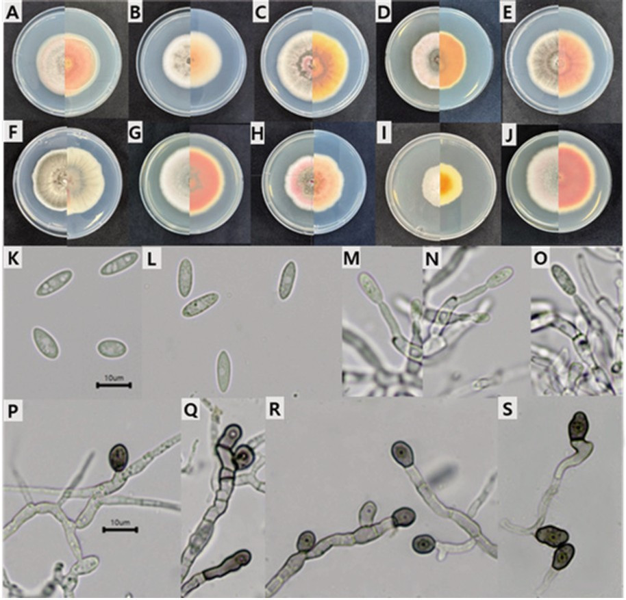
Fig. 1. Cultural and morphological characteristics of C. fioriniae isolates from apple. A–J: Seven-day-old colonies grown on potato dextrose agar showing variation in pigmentation from pinkish gray to olivaceous gray. K–L: Conidia; M–O: Conidiophores; P–S: Appressoria. Scale bars = 10 µm.
Table 1. Comparison of conidial and appressoria size of Colletotrichum fioriniae and C. orientalis associated with apple bitter rot in Korea and key references
| Species | Structure | Size range (µm) (L × W) | Mean size (µm) (L × W) | References |
|---|---|---|---|---|
| C. fioriniae | Conidia | (12.2–)14.3–15.7(–17.7) × (3.6–)4.3–5(–6.7) | 15.0 ± 1.1 × 4.7 ± 0.7 | This study |
| C. fioriniae | (10–)13.5–16.5(–19.5) × 4–5(–5.5) | 15.0 ± 1.6 × 4.5 ± 0.3 | [5] | |
| C. orientalis | (12.8–)14–16(–18.5) × (3.9–)4–5(–5.5) | 15.1 ± 1.2 × 4.5 ± 0.38 | [2] | |
| C. fioriniae | Appressoria | (5.8–)7.5–8.4(–11.2) × (4.4–)5.7–6.3(–8.0) | 8.4 ± 1.3 × 6.2 ± 0.9 | This study |
| C. fioriniae | (4.5–)7–11.5(–15.5) × (4–)4.5–7(–10.5) | 9.2 ± 2.2 × 5.6 ± 1.2 | [5] | |
| C. orientalis | (7–)8–9.5(–11.5) × (4.4–)5.5–6(–7.2) | 8.74 ± 0.99 × 5.84 ± 0.53 | [2] |
Maximum likelihood phylogenetic analysis of concatenated sequences from five loci (ITS, GAPDH, CHS-1, ACT, and TUB2) showed that all isolates obtained in this study clustered within the C. acutatum species complex and, more specifically, within the C. fioriniae clade (Fig. 2). The Korean isolates (GAC23017, GAC23054, GAC23072, GAC23079, GAC23083, GAC23084, GAC24004, GAC24005, GAC24003, and GAC24013) formed a well-supported clade with the ex-type strain C. fioriniae CBS 128517 and other reference strains, with high bootstrap support (> 90%). Furthermore, the Chinese strains previously described as C. orientalis (F10PGBYS1 and F10PGBYS08) were nested within the same lineage, indicating no phylogenetic separation between C. orientalis and C. fioriniae and thereby supporting their conspecificity. Pairwise sequence comparisons with CBS 128517 revealed only minor nucleotide differences, including 2 bp in GAPDH (0.79%), 2 bp in CHS-1 (0.71%), 2 bp in ACT (0.90%), and 3 bp in TUB2 (0.61%), whereas the ITS region was identical. All observed p-distance values were less than 1%, falling within the intraspecific variation reported for the C. fioriniae clade. These findings are consistent with those of Zhang et al. [6], who reported less than 1% divergence between C. orientalis and C. fioriniae across the same loci and argued that subtle differences in PHI-test recombination patterns alone are insufficient to justify recognizing C. orientalis as a distinct species. Consistent with these molecular data, recent taxonomic reassessments indicate that C. orientalis should be treated as a synonym of C. fioriniae because of overlapping morphological characteristics and limited multilocus sequence divergence [6]. These findings are in line with those of Sui et al. [10], who placed isolates from diverse forest plant hosts in China within the C. fioriniae clade, thereby further supporting the conspecificity of C. orientalis and C. fioriniae across multiple host species and geographic regions. They are also consistent with those of Shin et al. [7], who initially identified olive isolates as C. orientalis but subsequently reclassified them as C. fioriniae following evidence of conspecificity, designating C. fioriniae as the predominant causal agent of olive anthracnose in Korea.
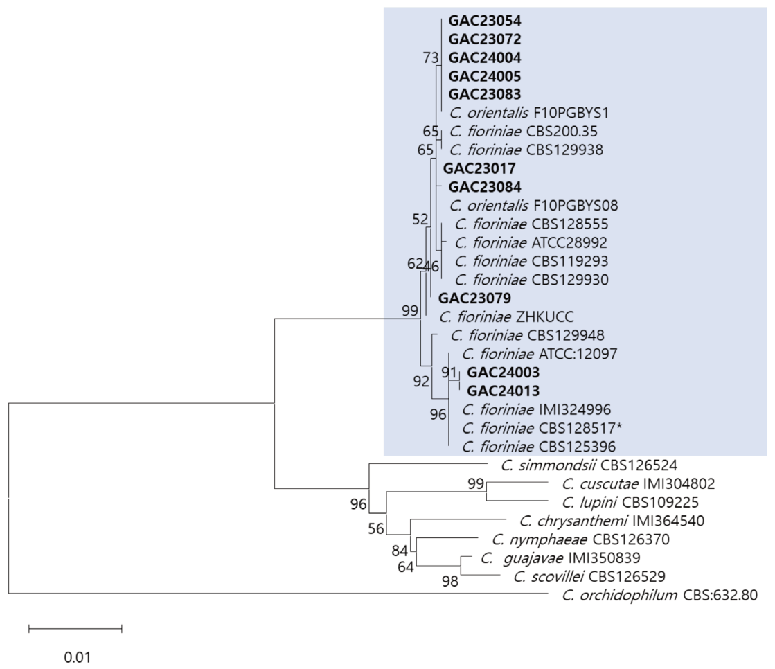
Fig. 2. Maximum likelihood phylogenetic tree of CASC based on concatenated multilocus sequences (internal transcribed spacer region, glyceraldehyde-3-phosphate dehydrogenase, chitin synthase 1, actin, and β-tubulin). Korean isolates (in bold) were clustered within the C. fioriniae clade together with the Chinese C. orientalis strains (F10PGBYS1, F10PGBYS08), indicating no phylogenetic separation. C. orchidophilum CBS 632.80 was used as the outgroup. Bootstrap support values (> 70%) are indicated at the nodes. Scale bar = 0.01 substitutions per site.
Pathogenicity assays were conducted on detached ʻFuji’ apple fruits under wounded and unwounded inoculation conditions to compare the virulence of isolates previously identified as C. orientalis and C. fioriniae. All tested isolates induced typical bitter rot symptoms, including brown lesions, on wounded fruits, whereas no symptoms were observed on unwounded or control fruits (Fig. 3C–H-1). The lesions that developed on wounded fruits were characteristic of apple bitter rot, and the inoculated fungus was successfully re-isolated from symptomatic tissues. The re-isolated cultures exhibited morphological characteristics consistent with those of the original isolates, thereby fulfilling Koch’s postulates. Mean lesion diameters on wounded ʻFuji’ fruits, calculated as the average of two perpendicular measurements per fruit, ranged from 6.90 to 11.82 mm among the five isolates tested. Isolates previously identified as C. orientalis (23017, 23084, and 24004) showed a mean (± SD) lesion diameter of 8.67 ± 1.31 mm, whereas isolates identified as C. fioriniae (24003 and 24013) exhibited a mean (± SD) lesion diameter of 9.45 ± 3.35 mm. Despite some isolate-to-isolate variation, there was no statistically significant difference in mean lesion diameter between isolates assigned to C. orientalis and C. fioriniae (independent-samples t-test, P > 0.05). The overlapping lesion size range and similar infection patterns indicate that the two groups exhibit comparable virulence and a common reliance on wound-mediated infection under the conditions tested. These pathogenicity results provide phenotypic evidence that the two nominal taxa are not distinguishable in their pathogenicity on apple fruits. When integrated with morphological characteristics and multilocus phylogenetic analyses, the pathogenicity data support the conclusion that Korean apple bitter rot isolates previously identified as C. orientalis should be regarded as C. fioriniae. This interpretation is consistent with recent molecular studies that report minimal sequence divergence between C. orientalis and C. fioriniae across multiple loci and place isolates assigned to these names within a single, well-supported clade [6,7]. It is also broadly consistent with current international nomenclatural frameworks, as C. orientalis is treated as a synonym of C. fioriniae in MycoBank, even though it is still listed as a current name in Index Fungorum, reflecting the transitional nature of this taxonomic revision [19,20]. Accordingly, the present study indicates that these Korean apple isolates are indistinguishable from C. fioriniae and should be treated as belonging to a single biological species, thereby providing a more coherent taxonomic basis for future work on apple bitter rot. Beyond taxonomic clarification, this study highlights several important directions for future research. Because species in the genus Colletotrichum can differ markedly in baseline fungicide sensitivity and resistance mechanisms, systematic monitoring of fungicide sensitivity and resistance in Korean C. fioriniae populations is warranted [21–24]. Establishing baseline sensitivities to the major fungicide classes used in apple production, together with investigations into the distribution and frequency of resistance-associated mutations and temporal trends in sensitivity, will provide a framework for designing effective resistance management strategies. The occurrence of C. fioriniae on apples and other hosts also suggests a broad ecological amplitude, underscoring the need for comparative studies across multiple hosts and regions to clarify host range, pathogenicity profiles, and environmental requirements for infection and disease development. Such work will provide useful information for improving disease prediction and for designing host- and species-specific management strategies that are consistent with the taxonomic framework proposed in this study.
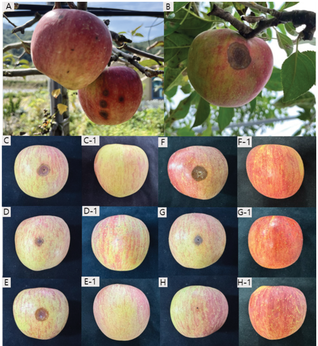
Fig. 3. Bitter rot symptoms on apple fruits and pathogenicity test results. A, B: Naturally infected ‘Fuji’ apple fruits showing typical brown, sunken bitter rot lesions in orchards. C–E: Wounded ‘Fuji’ fruits inoculated with three representative isolates previously identified as Colletotrichum orientalis (C: GAC23017, D: GAC23084, E: GAC24004), showing typical bitter rot lesions 7 days after incubation at 25°C. C-1–E-1: Corresponding unwounded fruits inoculated with the same isolates, showing no symptoms. F, G: Wounded ‘Fuji’ fruits inoculated with two representative isolates of C. fioriniae (F: GAC24003, G: GAC24013), showing typical bitter rot lesions. F-1, G-1: Corresponding unwounded fruits inoculated with the same isolates, showing no symptoms. H: Wounded control fruit treated with PDA plug only, showing no symptoms. H-1: Unwounded control fruit treated with PDA plug only, showing no symptoms under the same conditions. PDA: potato dextrose agar.
The authors declare that they have no potential conflicts of interest.
This study was supported by the Rural Development Administration Research Program (Project No. RS-2024-00396930).
1. Freeman S, Katan T, Shabi E. Characterization of Colletotrichum species responsible for anthracnose diseases of various fruits. Plant Dis 1998;82:596-605. https://doi.org/10.1094/pdis.1998.82.6.596
[DOI][PubMed]
2. Chen Y, Fu D, Wang W, Gleason ML, Zhang R, Liang X, Sun G. Diversity of Colletotrichum species causing apple bitter rot and Glomerella leaf spot in China. J Fungi 2022;8:740. https://doi.org/10.3390/jof8070740
[DOI][PubMed]
3. Nam YJ, Lee SY, Cho WD, Jung HY. First report of Colletotrichum godetiae causing postharvest bitter rot on apple in Korea. Plant Dis 2025;109:1591. https://doi.org/10.1094/PDIS-02-25-0416-PDN
[DOI]
4. Nam YJ, Lee SY, Cho WD, Jung HY. First report of anthracnose caused by Colletotrichum conoides on apple in Korea. Plant Dis 2025;109:496. https://doi.org/10.1094/PDIS-08-24-1735-PDN
[DOI]
5. Damm U, Cannon PF, Woudenberg JHC, Crous PW. The Colletotrichum acutatum species complex. Stud Mycol 2012;73:37-113. https://doi.org/10.3114/sim0010
[DOI][PubMed][PMC]
6. Zhang YX, Chen JW, Manawasinghe IS, Lin YH, Jayawardena RS, McKenzie EHC, Hyde KD, Xiang MM. Identification and characterization of Colletotrichum species associated with ornamental plants in Southern China. Mycosphere 2023;14:262-302. https://doi.org/10.5943/mycosphere/14/si2/5
[DOI]
7. Shin JH, Kim CH, Kim JE, Ahn JJ, Joa JH, Kim HL, Shin M, Lee SE, Gim MJ, Lee HJ, et al. Identification and characterization of Colletotrichum species associated with olive anthracnose in Korea. Mycobiology 2025;53:531-8. https://doi.org/10.1080/12298093.2025.2524882
[DOI][PubMed][PMC]
8. Jang HS, Hassan O, Chang T. First report of walnut anthracnose caused by Colletotrichum orientalis in South Korea. Plant Dis 2023;107:3633. https://doi.org/10.1094/PDIS-05-23-0833-PDN
[DOI]
9. Hassan O, Ju HY, Kim HS, Ryu H, Choi HW, Hong SK. First report of apple bitter rot caused by Colletotrichum orientalis in South Korea. Plant Dis 2025;109:2212. https://doi.org/10.1094/PDIS-03-25-0521-PDN
[DOI]
10. Sui XN, Guo MJ, Wang QT, Wang HQ, Zhou H, Huang XB, Yu ZF, Hou CL. Taxonomy and phylogeny of Colletotrichum on important forest plants in China. Mycosphere 2024;15:4569-743. https://doi.org/10.5943/mycosphere/15/1/19
[DOI]
11. Gelain J, Lykins S, Rosa PF, Soares AT, Dowling M, Schnabel G, May De Mio LL. Identification and fungicide sensitivity of Colletotrichum spp. from apple flowers and fruitlets in Brazil. Plant Dis 2023;107:1183-91. https://doi.org/10.1094/PDIS-01-22-0243-RE
[DOI][PubMed]
12. Dowling M, Peres N, Villani S, Schnabel G. Managing Colletotrichum on fruit crops: A “complex” challenge. Plant Dis 2020;104:2301-16. https://doi.org/10.1094/PDIS-11-19-2378-FE
[DOI][PubMed]
13. Usman HM, Tan Q, Karim MM, Adnan M, Yin WX, Zhu FX, Luo CX. Sensitivity of Colletotrichum fructicola and Colletotrichum siamense of peach in China to multiple classes of fungicides and characterization of pyraclostrobin-resistant isolates. Plant Dis 2021;105:3459-65. https://doi.org/10.1094/PDIS-04-21-0693-RE
[DOI][PubMed]
14. White TJ, Bruns TD, Lee SB, Taylor JW. Amplification and direct sequencing of fungal ribosomal RNA genes for phylogenetics. In: Innis MA, Gelfand DH, Sninsky JJ, White TJ, editors. PCR protocols: A guide to methods and applications. San Diego: Academic Press; 1990. p. 315-22. https://doi.org/10.1016/B978-0-12-372180-8.50042-1
[DOI][PubMed][PMC]
15. O’Donnell K, Cigelnik E. Two divergent intragenomic rDNA ITS2 types within a monophyletic lineage of the fungus Fusarium are nonorthologous. Mol Phylogenet Evol 1997;7:103-16. https://doi.org/10.1006/mpev.1996.0376
[DOI][PubMed]
16. Templeton MD, Rikkerink EH, Solon SL, Crowhurst RN. Cloning and molecular characterization of the glyceraldehyde-3-phosphate dehydrogenase-encoding gene and cDNA from the plant pathogenic fungus Glomerella cingulata. Gene 1992;122:225-30. https://doi.org/10.1016/0378-1119(92)90055-T
[DOI][PubMed]
17. Carbone I, Kohn LM. A method for designing primer sets for speciation studies in filamentous ascomycetes. Mycologia 1999;91:553-6. https://doi.org/10.1080/00275514.1999.12061051
[DOI]
18. Kumar S, Stecher G, Li M, Knyaz C, Tamura K. MEGA X: Molecular evolutionary genetics analysis across computing platforms. Mol Biol Evol 2018;35:1547-9. https://doi.org/10.1093/molbev/msy096
[DOI][PubMed]
19. MycoBank. Colletotrichum orientalis Dandan Fu & G.Y. Sun [Internet]. Utrecht: Westerdijk Fungal Biodiversity Institute [cited 2026 Aug 18]. Available from https://www.mycobank.org/page/Name%20details%20page/521273
20. Index Fungorum. Colletotrichum orientalis Dandan Fu & G.Y. Sun [Internet]. Kew: Royal Botanic Gardens; 2026 [cited 2026 Aug 18]. Available from https://www.indexfungorum.org/names/NamesRecord.asp?RecordID=845061
21. Ishii H, Zhen F, Hu M, Li X, Schnabel G. Efficacy of SDHI fungicides, including benzovindiflupyr, against Colletotrichum species. Pest Manag Sci 2016;72:1844-53. https://doi.org/10.1002/ps.4216
[DOI][PubMed]
22. Cao X, Xu X, Che H, West JS, Luo D. Distribution and fungicide sensitivity of Colletotrichum species complexes from rubber tree in Hainan, China. Plant Dis 2017;101:1774-80. https://doi.org/10.1094/PDIS-03-17-0352-RE
[DOI][PubMed]
23. Johnson KA, Douglas RK, Bradshaw MJ, Brannen PM, Jurick WM, Villani SM. Colletotrichum species causing Glomerella leaf spot and apple bitter rot in the southeastern United States exhibit disparities in relative frequency, morphological phenotype, and quinone outside inhibitor sensitivity. Plant Dis 2025;109:579-92. https://doi.org/10.1094/PDIS-05-24-1006-RE
[DOI][PubMed]
24. Cortaga CQ, Cordez BWP, Dacones LS, Balendres MAO, Dela Cueva FM. Mutations associated with fungicide resistance in Colletotrichum species: A review. Phytoparasitica 2023;51:569-92. https://doi.org/10.1007/s12600-023-01063-0
[DOI]