
Jae Sung Lee1, Yoo-Kyeong Lee2, Bora Kim3, Seong-Jin Lee4, Young-Joon Choi2,*, and Hyeon-Dong Shin5
1Department of Agricultural Biology, National Institute of Agricultural Sciences, Rural Development Administration, Wanju 55365, Korea
2Department of Biological Science, Kunsan National University, Gunsan 54150, Korea
3Climate Change and Carbon Research Team, National Institute of Ecology, Seocheon 33657, Korea
4Department of Plant Quarantine, Animal and Plant Quarantine Agency, Gimcheon 39660, Korea
5Division of Environmental Science and Ecological Engineering, Korea University, Seoul 02842, Korea
*Corresponding author: yjchoi@kunsan.ac.kr
Korean Journal of Mycology (Kor J Mycol) 2026 September, Volume 54, Issue 3, pages 231-236.
https://doi.org/10.4489/kjm.2026.54.3.2
Received on June 01, 2026, Revised on July 31, 2026, Accepted on August 18, 2026, Published on September 30, 2026.
© 2026 THE KOREAN SOCIETY OF MYCOLOGY.
This is an Open Access article distributed under the terms of the Creative Commons Attribution Non-Commercial License (http://creativecommons.org/licenses/by-nc/4.0/) which permits unrestricted non-commercial use, distribution, and reproduction in any medium, provided the original work is properly cited.
Asian fawn lily, Liliaceae, Phylogenetic analysis
Erythronium japonicum Decne., the Asian fawn lily, is a spring-flowering Liliaceae species native to northeastern China, Japan, Korea, and the Russian Far East [1]. This plant is widely cultivated as a popular ornamental plant in Korea owing to its large and attractive flowers. In addition, E. japonicum has been traditionally used as a medicinal plant in Korea, and several studies have investigated its phytochemical constituents [2,3].
In April of 2022 and 2023, E. japonicum plants at Pocheon (37°45′24″N, 127°09′46″E), Wanju (35°58′24″N, 127°18′12″E), and Jinan (35°45′44″N, 127°24′17″E), Korea, exhibited rust symptoms, with disease incidence reaching up to 80%. Symptoms appeared as yellow to brown, vein-limited spots (Fig. 1A and 1B) on the upper leaf surfaces, with orange-colored spermogonia, aecia (Fig. 1C), and black telia forming mainly on the corresponding lower surfaces (Fig. 1J and 1K) but occasionally on the upper surfaces and petioles. Five representative samples were deposited in the Kunsan National University Herbarium (KSNUH1816, 1817) and Korea University Herbarium (KUS-F32715, F33601, and F33614).
For morphological characterization, the infected leaf tissues were examined using a stereomicroscope (M205C, Leica, Wetzlar, Germany) and a differential interference contrast light microscope (Axio Imager 2, Carl Zeiss, Oberkochen, Germany). The fine surface structures were further investigated using a scanning electron microscope (S-4800 with EDS, Hitachi, Tokyo, Japan). Spermogonia were amphigenous on the leaf blades and petioles, scattered, round, pale yellow, conical-shaped, and present among the aecia (Fig. 1D). Aecia were amphigenous or cauligenous, densely grouped, light yellow to orange, cup-shaped with peridia, and measured 350–520 μm (av. 440 μm) in diameter (Fig. 1E and 1F). Aeciospores were globose to subglobose, polygonal, pale yellow, and measured 20–26×18–23 μm (av. 23.2×20.9 μm) (Fig. 1G and 1H), with a verrucose and hyaline wall (Fig. 1I). No uredinia were observed. Telia were amphigenous, elongated, and measured 390–760 μm (av. 480 μm) in diameter (Fig. 1L). Teliospores were one-celled, ovoid, dark brown, measured 25–37×20–25 μm (av. 30.3×23.0 μm), with a short pedicel (Fig. 1M). The teliospore walls had long ridges that ran lengthwise (Fig. 1N). These morphological characteristics corresponded well with those of Uromyces erythronii (DC.) Pass. [4].
To further verify the morphological identification, genomic DNA was extracted from aeciospores and teliospores collected from naturally infected leaves using the MagListo 5M Plant Genomic DNA Extraction Kit (Bioneer, Daejeon, Korea). The internal transcribed spacer (ITS) and large subunit (LSU) rDNA regions were amplified and sequenced using the primers ITS5-u/ITS4rust [5,6] and LRust1R/ LRust3 [6], respectively. The PCR products were purified using AccuPrep® PCR/Gel Purification Kit (Bioneer, Daejeon, Korea) and sequenced by Macrogen sequencing service (Macrogen, Seoul, Korea) with the primers used for amplification. The resulting sequences were deposited in GenBank (accession nos. PV545142.1–PV545146.1 for ITS, PV545147.1–PV545151.1 for LSU). The BLASTn results showed that the ITS sequences were identical to three sequences of U. erythronii (MH205917.1, MH205916.1, and LC203755.1), and the LSU sequences matched those of U. erythronii from E. japonicum in Japan (LC203755.1). Phylogenetic trees were constructed by the maximum-likelihood method using MEGA7 [7] with the default settings of the program and the Tamura–Nei model. The robustness of the individual branches was estimated by bootstrapping 1,000 replicates.
Phylogenetic analyses based on both the ITS and LSU sequences consistently placed all Korean samples within the U. erythronii clade (Fig. 2A and 2B). In the ITS tree (Fig. 2A), the Korean collections (KSNUH1816, 1817, KUS-F33614, F32715, and F33601) grouped together with authentic U. erythronii reference sequences obtained from E. japonicum in Japan (LC203755.1) and E. dens-canis in Romania (MH205916.1) and Croatia (MH205917.1), forming a highly supported monophyletic clade with a high bootstrap value (BS) of 99%. This clade was clearly separated from other Uromyces species, including U. trifolii-repentis, U. viciae-fabae, U. beticola, U. durus, U. gageae, U. muscari, U. transversalis, and U. japonicus. In the LSU tree (Fig. 2B), all Korean isolates clustered with the Japanese reference sequence of U. erythronii (LC203755.1), with strong bootstrap support (BS = 96%). The topology of the LSU tree was congruent with that of the ITS tree, confirming that the Korean specimens were conspecific with U. erythronii.
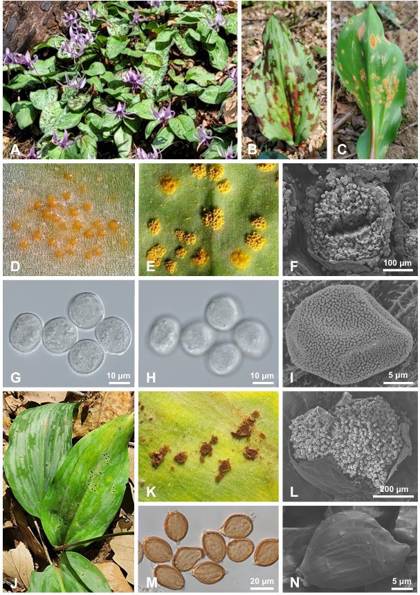
Fig. 1. Rust disease caused by Uromyces erythronii on Erythronium japonicum in South Korea. A, Rust symptoms on E. japonicum in a forest. B, Brown to yellow chlorotic spots on the upper surface of the infected leaf. C, Aecia on the lower leaf surface. D, Spermogonia. E, Aecia. F, Aecium and aeciospores observed using an SEM. G and H, Aeciospores observed using a DIC microscope. I, Verrucose wall ornamentation of an aeciospore under an SEM. J, Telia on the infected leaves. K, Telia. L, Telium and teliospores under an SEM. M, Teliospores under a DIC microscope. N, Wall ornamentation of a teliospore under an SEM. SEM: scanning electron microscope; DIC: differential interference contrast.
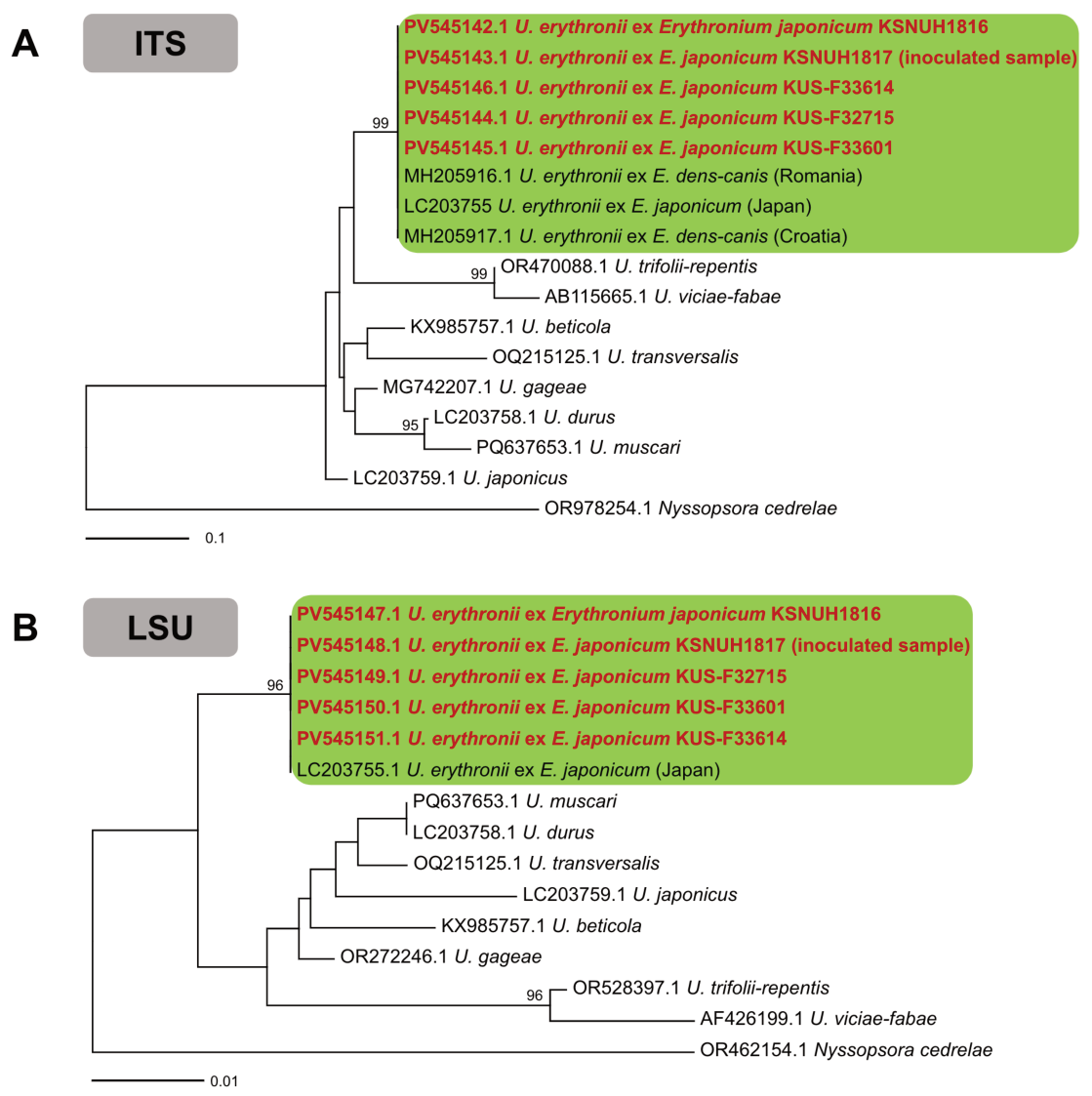
Fig. 2. Maximum-likelihood phylogenetic trees of Uromyces species based on ITS (A) and LSU (B) rDNA sequences. Bootstrap support values higher than 60% are shown above the branches. Korean samples are highlighted in bold red. ITS: internal transcribed spacer; LSU: large subunit.
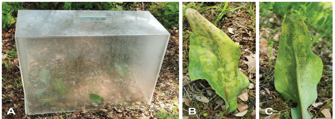
Fig. 3. Pathogenicity test of Uromyces erythronii on Erythronium japonicum. A, Erythronium japonicum plants inoculated with aeciospores and maintained in a plastic chamber. B and C, Telia developed on the upper (B) and lower (C) leaf surfaces two weeks after inoculation.
Pathogenicity was assessed by inoculating three healthy E. japonicum plants with aeciospores (1.1×10⁶ spores/mL) from naturally infected leaves. Three uninoculated plants served as controls. All plants were maintained in a plastic chamber at 15–20°C with high humidity (Fig. 3A). After two weeks, all inoculated plants developed dark brown telia on their leaves (Fig. 3B and 3C), whereas the control plants remained symptomless. Formation of the telial stage from aeciospore inoculation was consistent with the life cycle of U. erythronii previously described by Fukuda and Nakamura [8]. The pathogen on the inoculated plants was confirmed as U. erythronii by sequencing, thus fulfilling Koch’s postulates.
To our knowledge, this is the first report of rust disease caused by U. erythronii on E. japonicum in South Korea; however, it has previously been recorded only in Japan and North Korea [9,10]. Uromyces erythronii has been reported on Amana, Erythronium, and Lilium species (Liliaceae) in Asia (China and Japan), as well as several European countries (Bulgaria, Croatia, France, Germany, Greece, Hungary, Italy, Romania, Switzerland, Spain, and Ukraine) [11,12]. Considering the high disease incidence and severity observed in Korea, U. erythronii may threaten the cultivation and conservation of E. japonicum.
The authors declare that they have no potential conflict of interest.
This study was supported by grants from the Animal and Plant Quarantine Agency (I-1543086-2025-28-01 & Z-1543086-2025-28-01), Korea.
1. Mathew B. Plate 411. Erythronium japonicum: Liliaceae. Curtis’s Bot Mag 2001;18(2):68-73. https://doi.org/10.1111/1467-8748.00290
[DOI]
2. Lee MS, Lim SC, Park HJ. Phthalate ester and flavonoids isolated from leaves of Erythronium japonicum. Korean J Med Crop Sci 1994;2:67-72.
3. Park YJ, Ku YG, Kang NR, Cho JY, Heo BG. Amino acid contents and various physiological activities of Erythronium japonicum. J People Plants Environ 2015;18:345-51. https://doi.org/10.11628/ksppe.2015.18.5.345
[DOI]
4. Tykhonenko YY, Sytschak NN, Kagalo AA, Orlov OO. New records of Uromyces erythronii (Pucciniales) from Ukraine. Ukr Bot J 2017;74:184-8. https://doi.org/10.15407/ukrbotj74.02.184
[DOI]
5. Pfunder M, Schürch S, Roy BA. Sequence variation and geographic distribution of pseudoflower-forming rust fungi (Uromyces pisi s. lat.) on Euphorbia cyparissias. Mycol Res 2001;105:57-66. https://doi.org/10.1017/S0953756200003208
[DOI]
6. Beenken L, Zoller S, Berndt R. Rust fungi on Annonaceae II: The genus Dasyspora Berk. & M.A. Curtis. Mycologia 2012;104:659-81. https://doi.org/10.3852/11-068
[DOI][PubMed]
7. Kumar S, Stecher G, Tamura K. MEGA7: Molecular evolutionary genetics analysis version 7.0 for bigger datasets. Mol Biol Evol 2016;33:1870-4. https://doi.org/10.1093/molbev/msw054
[DOI][PubMed]
8. Fukuda T, Nakamura S. Biotic interaction between a rust fungus, Uromyces erythronii Pass., and its host plant, Erythronium japonicum Decne. (Liliaceae). Plant Species Biol 1987;2:75-83. https://doi.org/10.1111/j.1442-1984.1987.tb00034.x
[DOI]
9. Hiratsuka N. Uredinales collected in Korea V. Bot Mag (Tokyo) 1942;56:53-61. https://doi.org/10.15281/jplantres1887.56.53
[DOI]
10. Hiratsuka N, Sato S, Katsuya K, Kakishima M, Hiratsuka Y, Kaneko S, Ono Y, Sato T, Harada Y, Hiratsuka T, et al. The rust flora of Japan. Tsukuba: Tsukuba Shuppankai; 1992.
11. Nagy T, Pfliegler WP, Takács A, Tökölyi J, Molnár VA. Distribution, infection rates and DNA barcoding of Uromyces erythronii (Pucciniaceae), a parasite of Erythronium (Liliaceae) in Europe. Willdenowia 2019;49:13-20. https://doi.org/10.3372/wi.49.49103
[DOI]
12. Farr DF, Rossman AY. Fungal databases, U.S. National Fungus Collections [Internet]. Beltsville (MD): ARS, USDA; 2025 [cited 2025 Apr 10]. Available from https://nt.ars-grin.gov/fungaldatabases/