
Kyoung-Mo Koo1, Gyo-Seon Shin2, Ji-Hyun Park1,2,3,*, and Hyeon-Dong Shin3,4
1Department of Forest Resources, Kookmin University, Seoul 02707, Korea
2Forest Carbon Graduate School, Kookmin University, Seoul 02707, Korea
3Department of Forestry, Environment, and Systems, Kookmin University, Seoul 02707, Korea
4Division of Environmental Science and Ecological Engineering, Korea University, Seoul 02841, Korea
*Corresponding author: jhpark10@kookmin.ac.kr
Korean Journal of Mycology (Kor J Mycol) 2026 September, Volume 54, Issue 3, pages 237-249.
https://doi.org/10.4489/kjm.2026.54.3.3
Received on July 22, 2026, Revised on August 11, 2026, Accepted on August 18, 2026, Published on September 30, 2026.
© 2026 THE KOREAN SOCIETY OF MYCOLOGY.
This is an Open Access article distributed under the terms of the Creative Commons Attribution Non-Commercial License (http://creativecommons.org/licenses/by-nc/4.0/) which permits unrestricted non-commercial use, distribution, and reproduction in any medium, provided the original work is properly cited.
Identification, Phylogeny, Physiology, Prunus persica, Taphrinaceae
Taphrina (Taphrinaceae, Taphrinales) comprises dimorphic ascomycetous fungi that infect a broad range of vascular plant hosts [1–4]. During their sexual stage, the fungus develops dikaryotic hyphae within living host tissues, intercellularly, subcuticularly, or under the epidermis, and produces naked asci on the surface of diseased organs. Infection causes hypertrophy and deformation, resulting in symptoms such as fruit pockets, leaf blotch, leaf curl, leaf gall, tongue gall, and witches’ brooms [1–4]. In the asexual stage, Taphrina species exhibit yeast-like growth, reproducing by budding, and can be cultured on artificial media under laboratory conditions [1–4].
Traditionally, species in Taphrina have been delineated based on host association, disease symptoms, and morphological characteristics, including the size and shape of asci, ascospores, and stalk cells [1–3]. More recently, molecular phylogenetic analysis has become an essential tool for species identification and classification. The internal transcribed spacer (ITS) region of the ribosomal RNA (rRNA) gene, the D1 and D2 domains of the 26S rRNA gene, and the mitochondrial small subunit ribosomal RNA (rns) gene are the molecular markers most commonly used for phylogenetic studies of Taphrina, as they provide improved species delimitation and taxonomic resolution [4–8]. Based on these combined morphological and molecular data, approximately 30 species are currently recognized in the genus [4,9].
Taphrina deformans, the causal agent of leaf curl on almond (Prunus dulcis (Mill.) D.A. Webb) and peach (P. persica (L.) Batsch), currently has a worldwide distribution [2,4]. However, in Korea, records of peach leaf curl are based primarily on historical specimens collected prior to the advent of molecular analysis, and only limited morphological examinations have been performed; thus, the pathogen’s identity has not been confirmed using a modern polyphasic approach [10].
Peach is a major fruit crop cultivated in countries worldwide, including Korea, where its production ranks fourth among domestic fruit crops [11,12]. In addition to its agricultural importance, peach-based agroforestry systems have been recognized for their potential in degraded land restoration, soil erosion control, slope stabilization, and the enhancement of ecosystem services [13,14]. However, peach is highly susceptible to peach leaf curl caused by T. deformans. The disease reduces photosynthetic activity and induces premature defoliation, and severe outbreaks may affect up to 90% of the tree’s shoots, substantially reducing fruit production and tree vigor [15,16]. In Korea, peach leaf curl is regularly observed in peach orchards and is recognized as one of the major diseases affecting peach production [17].
The objective of this study was to definitively identify the fungus associated with peach leaf curl through disease symptom observations and morphological characterization, together with molecular phylogenetic analyses. In addition, the physiological characteristics of the isolates were investigated to provide further taxonomic evidence for the classification of these ascomycetous yeasts.
Leaves of Prunus persica exhibiting curls were collected from orchards in Jeonju (35°52′36″N, 127°06′38″E); Osan (37°09′57″N, 127°03′29″E); Dongdaemun-gu, Seoul (37°35′35″N, 127°02′40″E ); Wanju (35°49′35″N, 127°01′48″E); Jongno-gu, Seoul (37°34′39″N, 126°59′35″E); and Seongbuk-gu, Seoul (37°36′40″N, 127°00′02″E). After collection, samples were immediately transported to the laboratory under refrigerated conditions, and specimen preparation, microscopic observation, and fungal isolation were conducted aseptically within 6 hr to minimize changes in sample condition. Voucher specimens were deposited in the Korea University Mycological Herbarium (KUS-F) (Table 1).
Table 1. Voucher specimen numbers, culture collection information, and NCBI GenBank accession numbers for the Taphrina isolates examined in this study
| KUS-F voucher no. | Locality | Collection date | KACC accession no. | NCBI GenBank accession no. ITS | NCBI GenBank accession no. D1/D2 | NCBI GenBank accession no. rns |
|---|---|---|---|---|---|---|
| KUS-F35004 | Jeonju-si, Jeollabuk-do | 6 May 2021 | Not deposited | Not submitted | ||
| KUS-F35005 | Osan-si, Gyeonggi-do | 13 May 2021 | KACC 411353 | PZ626205 | PZ626210 | PZ630116 |
| KUS-F35006 | Dongdaemun-gu, Seoul | 1 Jun 2021 | Not deposited | Not submitted | ||
| KUS-F35007 | Wanju-gun, Jeollabuk-do | 29 Apr 2024 | KACC 411354 | PZ626206 | PZ626209 | PZ630115 |
| KUS-F35008 | Jongno-gu, Seoul | 28 May 2024 | Not deposited | Not submitted | ||
| KUS-F35009 | Seongbuk-gu, Seoul | 4 Jun 2025 | KACC 411352 | PZ626208 | PZ626211 | PZ630117 |
KUS-F: Korea University Mycological Herbarium; KACC: Korean Agricultural Culture Collection; NCBI: National Center for Biotechnology Information; ITS: Internal transcribed spacer region of the rRNA gene; D1/D2: D1/D2 domains of 26S rRNA gene; rns: Mitochondrial small subunit ribosomal RNA gene.
Pure cultures were obtained using a spore-drop isolation method. Approximately 3 mm² sections bearing hymenium tissue were cut from the margins of leaves with a sterile scalpel and affixed to the inner side of a Petri dish lid. Spores were then allowed to fall onto glucose–peptone–yeast extract (GPY) agar (2% glucose, 1% peptone, 0.5% yeast extract, and 2% agar) [18]. The lid was rotated every 2 hr, and the new position of the tissue section was marked to identify the corresponding spore deposition site. After incubation at 21℃ for 5 days, individual colonies appearing at the marked sites were transferred to yeast malt (YM) agar using a sterile loop. Samples from each isolate were preserved at −80℃ in 20% glycerol, and three representative isolates were deposited in the Korean Agricultural Culture Collection (KACC) (Table 1).
Morphological characteristics were examined using freshly collected tissues. Fungal structures, including ascogenous cells, asci with stalk cells, ascospores, and blastospores, were characterized under a compound light microscope (Olympus BX53-32XDIC equipped with a DP74 camera, Olympus, Tokyo, Japan). For each morphological feature, at least 50 individual structures were measured.
Genomic DNA was extracted from 7-day-old cultures grown on YM agar using the Quick-DNA Fungal/Bacterial Miniprep Kit (Zymo Research, Irvine, CA, USA). The ITS region, D1/D2 domains of the 26S rRNA gene, and rns gene were amplified with the primer pairs ITS1/ITS4, NL1/NL4, and SSU1/ SSU5, respectively (Table 2). Polymerase chain reactions were carried out using PCR Master Mix, 2× (Promega, Madison, WI, USA), and the amplified products were sequenced through a DNA sequencing service (Cosmogenetech, Seoul, Korea). The obtained sequences were edited using Chromas version 2.6.6 (Technelysium Pty Ltd., South Brisbane, Australia) and aligned in BioEdit version 7.2.5 [21]. The assembled ITS region, D1/D2 domain, and rns gene sequences were deposited in the National Center for Biotechnology Information (NCBI) GenBank database (Table 1). Sequence similarity searches were conducted against the GenBank database using BLAST [22].
For phylogenetic analysis, a total of 60 sequences representing 38 Taphrina species were obtained from the NCBI GenBank and National Agriculture and Food Research Organization (Japan) Genebank databases, with Protomyces inouyei (HA 1353) selected as the outgroup [4,8]. The ITS, D1/D2, and rns sequences were concatenated in MEGA 11 [23] and aligned using the MAFFT online service version 7.0 [24]. Candidate nucleotide substitution models were evaluated with jModelTest version 2.1.10 [25], and the best-fitting model was chosen based on the Akaike information criterion [26]. A maximum likelihood phylogenetic analysis was conducted using PhyML version 3.0 [27], with branch support assessed using 1,000 bootstrap resamples.
Table 2. Primer pairs used to amplify the ITS, D1/D2, and rns regions
| Primer | Primer sequence (5′–3′) | Target region | Reference |
|---|---|---|---|
| ITS1 | TCCGTAGGTGAACCTGCGG | ITS region (of rDNA) | [19] |
| ITS4 | TCCTCCGCTTATTGATATGC | ||
| NL1 | GCATATCAATAAGCGGAGGAAAAG | D1/D2 domain (of LSU rDNA) | [20] |
| NL4 | GGTCCGTGTTTCAAGACGG | ||
| SSU1 | GTGCCAGCAGTCGCGGTAATAC | rns gene (of mtDNA) | [7] |
| SSU5 | CAGGTTCCCCTACAGTAACTGTATTTCAACTT |
ITS: internal transcribed spacer; rDNA: ribosomal DNA; D1/D2: D1/D2 domains of the 26S rRNA gene; LSU: large subunit; rns: mitochondrial small subunit ribosomal RNA gene; mtDNA: mitochondrial DNA.
The physiological characteristics of the strains were investigated according to the standard procedures described by Kurtzman et al. [18]. Carbon assimilation was evaluated for 41 carbon sources in 16 × 125 mm test tubes containing 5 mL of liquid medium, and nitrogen assimilation was assessed for 9 nitrogen sources on solid media in 90 × 15 mm Petri dishes. All assays were performed at 21℃, with 21-day incubation periods for carbon assimilation tests and 14-day incubation periods for nitrogen assimilation tests. Urea hydrolysis and tolerance to 0.01% cycloheximide were examined in liquid medium using 16 × 125 mm test tubes. In addition, the diazonium blue B (DBB) color reaction and growth at 20, 25, and 30℃ on YM agar were evaluated. Lastly, the production of starch-like polysaccharides was assessed using both liquid and solid media. All tests were carried out independently in triplicate to ensure the reproducibility of the results (Fig. S1).
During field surveys conducted across six regions of South Korea, we detected leaf curl symptoms on Prunus persica in all six regions beginning in mid-May (Fig. 1A). Symptoms initially appeared as small reddish swellings on young leaves (Fig. 1B). As the disease progressed, the affected tissues became enlarged and thickened, resulting in leaf curl and distortion, with the development of an amphigenous white hymenial layer (Fig. 1C). In advanced stages, the affected tissues turned brown and became necrotic, and in severe cases the symptoms extended over nearly the entire leaf (Fig. 1D).
The sexual state developed on diseased leaves as a hymenial layer (Fig. 1E–G). Ascogenous cells, 10–20 × 6.5–13 μm in size, that developed into immature asci were observed on the epidermal surface (Fig. 1E). As development progressed, a mature naked ascus layer formed on both sides of the infected leaf lesions (Fig. 1F, G). Asci were cylindrical with a rounded apical region, hyaline, and 22–38 × 9–15 μm in size (n = 50) (Fig. 1H). Each ascus contained eight biseriately arranged ascospores, and budding within the ascus was frequently observed (Fig. 1H). Stalk cells were not observed in the present study. Ascospores were hyaline, globose to subglobose, and ovate or obovate, measuring 6–10 × 5–7 μm (n = 50) (Fig. 1I). After 7 days of incubation on YM agar at 21℃, an asexual, blastospore-producing yeast-like state derived from ascospores was observed. Blastospores were subglobose, ovate, or elliptical and reproduced by budding (Fig. 1J). On YM agar, cultures produced yeast-like growth along streaking lines, forming smooth, butyrous colonies with a yellowish-cream to pinkish-cream coloration (Fig. 1K).
The ITS, D1/D2, and rns sequences were identical among all Korean isolates. BLAST searches against the NCBI GenBank database indicated that these isolates’ ITS and D1/D2 domain regions shared 100% sequence identity with those of the type strain of T. deformans, CBS 356.35T, isolated from Prunus persica in the Netherlands (accession no. NR_160071 for ITS and accession no. AF492038 for D1/D2 domain).
For the ML phylogenetic analysis, the final alignment consisted of 60 sequences and 2,315 characters, comprising the concatenated ITS, D1/D2, and rns sequences, and the best-fit nucleotide substitution model was GTR+I+G. The phylogeny showed that all Taphrina deformans isolates, including the Korean strains, the type strain CBS 356.35T, and additional reference strains (CBS 355.35, NRRL-T470, PYCC 5894, and MAFF 242963), formed a single, well-supported clade (Fig. 2).
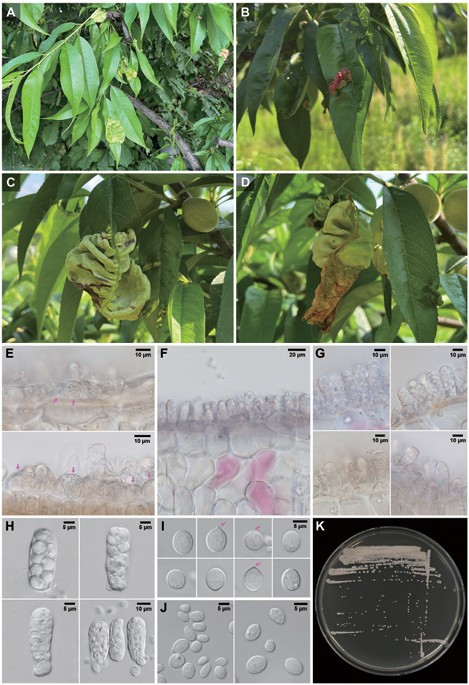
Fig. 1. Images showing the symptomatic features and morphological characteristics of the leaf curl pathogen on Prunus persica. A–D: leaf curl symptoms on P. persica infected with T. deformans in mid-May; E: ascogenous cells observed on the surface of infected leaves (arrows); F and G: cross-sectional views of the naked ascus layer; H: asci; I: ascospores, with budding indicated by arrows; J: blastospores; and K: colonies grown on yeast malt agar at 21℃ for 7 days.
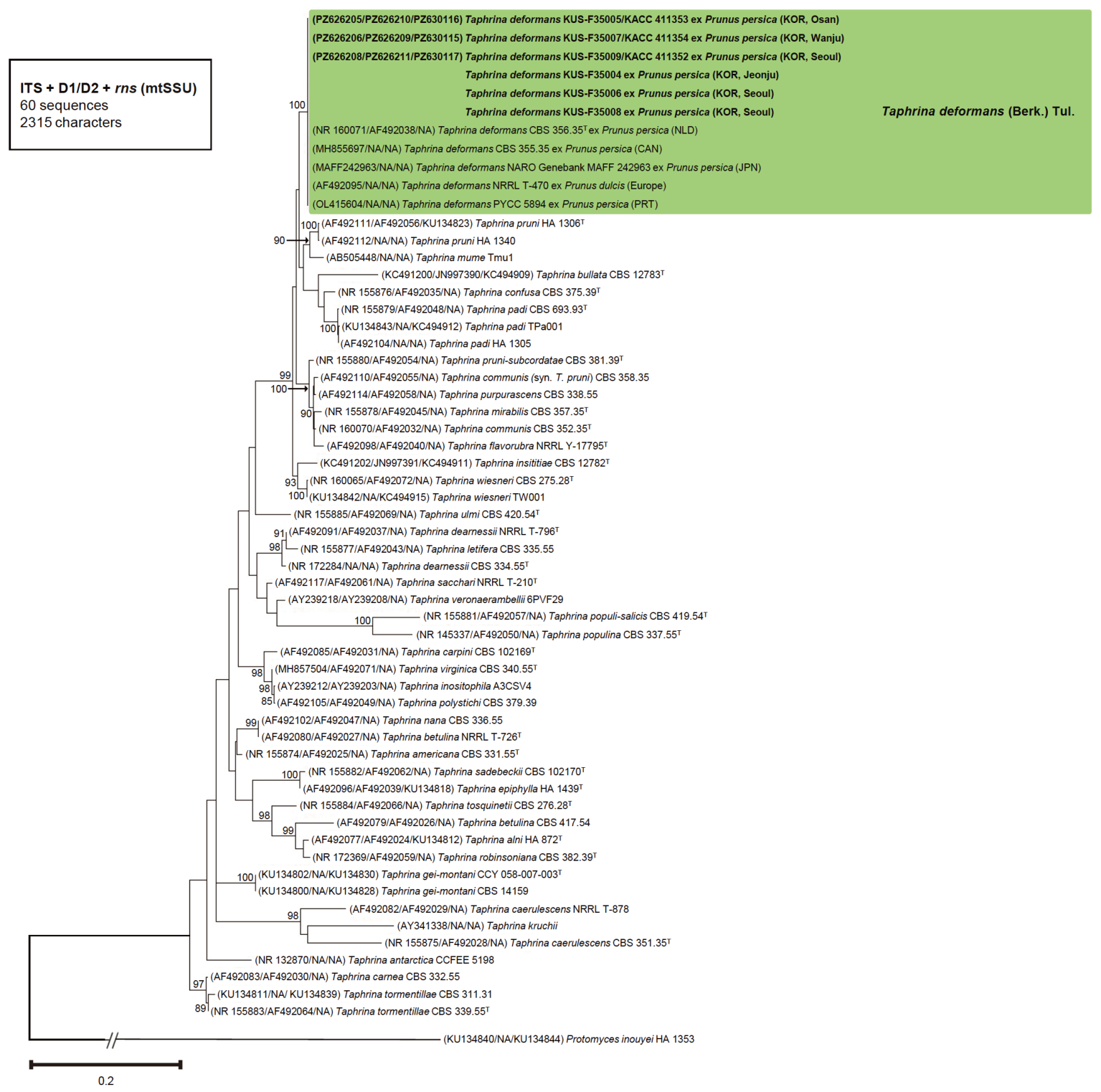
Fig. 2. Phylogenetic tree of Taphrina deformans (green box) and related Taphrina species, inferred from the concatenated ITS, D1/D2, and rns sequences of 38 Taphrina species using the maximum likelihood method based on the GTR+I+G nucleotide substitution model in PhyML version 3.0. Bootstrap support values > 70%, obtained from 1,000 replicates, are shown above the corresponding nodes. The isolates obtained in this study are shown in bold, and type strains are indicated by a superscript T. The scale bar represents 0.2 nucleotide substitutions per site, and “NA” indicates that the sequence data were unavailable. ITS: internal transcribed spacer; D1/D2: D1/D2 domains of the 26S rRNA gene; rns: mitochondrial small subunit ribosomal RNA gene; GTR+I+G: general time reversible model with a proportion of invariable sites and gamma-distributed rate heterogeneity.
Out of 41 tested carbon compounds, 13 were assimilated (+) by all three assessed strains: D-glucose, D-xylose, D-arabinose, sucrose, trehalose, cellobiose, salicin, raffinose, glycerol, xylitol, D-glucitol, D-mannitol, and fructose. The remaining 28 were not (−): D-galactose; L-sorbose; D-glucosamine; D-ribose; L-arabinose; L-rhamnose; maltose; α-methyl-D-glucoside; arbutin; melibiose; lactose; melezitose; inulin; soluble starch; erythritol; ribitol; galactitol; myo-inositol; D-glucono-1,5-lactone; D-glucuronate; DL-lactate; succinate; citrate; methanol; ethanol; N-acetyl-D-glucosamine; L-malate; and hexadecane. Out of nine tested nitrogen compounds, three, nitrate (potassium), D-glucosamine, and ammonium sulfate, were assimilated (+), whereas nitrite (sodium), ethylamine, L-lysine, cadaverine, creatinine, and N-acetyl-D-glucosamine were not (−). No growth was observed in the vitamin-free medium or the medium containing 0.01% cycloheximide (−), and the DBB reaction was negative (−). In temperature growth tests, growth occurred at 20℃ and 25℃ but not at 30℃ (Table 3).
Table 3. Comparison of physiological characteristics between Korean isolates and reference strains of Taphrina deformans
| Physiological characteristic – C: Carbon source – N: Nitrogen source | Previous study [4] Taphrina deformans | Taphrina deformans (Korean isolates) KACC 411352 | This study KACC 411353 | KACC 411354 |
|---|---|---|---|---|
| C1 D-Glucose | + | + | + | + |
| C2 D-Galactose | − | − | − | − |
| C3 L-Sorbose | v | − | − | − |
| C4 D-Glucosamine | − | − | − | − |
| C5 D-Ribose | − | − | − | − |
| C6 D-Xylose | + | + | + | + |
| C7 L-Arabinose | − | − | − | − |
| C8 D-Arabinose | − | + | + | + |
| C9 L-Rhamnose | − | − | − | − |
| C10 Sucrose | v | + | + | + |
| C11 Maltose | − | − | − | − |
| C12 Trehalose | v | + | + | + |
| C13 α-methyl-D-glucoside | − | − | − | − |
| C14 Cellobiose | + | + | + | + |
| C15 Salicin | + | + | + | + |
| C16 Arbutin | nd | − | − | − |
| C17 Melibiose | − | − | − | − |
| C18 Lactose | − | − | − | − |
| C19 Raffinose | − | + | + | + |
| C20 Melezitose | − | − | − | − |
| C21 Inulin | − | − | − | − |
| C22 Soluble starch | v | − | − | − |
| C23 Glycerol | v | + | + | + |
| C24 Erythritol | − | − | − | − |
| C25 Ribitol | − | − | − | − |
| C26 Xylitol | v | + | + | + |
| C27 D-Glucitol | + | + | + | + |
| C28 D-Mannitol | + | + | + | + |
| C29 Galactitol | − | − | − | − |
| C30 myo-Inositol | − | − | − | − |
| C31 D-Glucono-1,5-lactone | v | − | − | − |
| C32 D-Glucuronate | − | − | − | − |
| C33 DL-Lactate | − | − | − | − |
| C34 Succinate | v | − | − | − |
| C35 Citrate | − | − | − | − |
| C36 Methanol | − | − | − | − |
| C37 Ethanol | v | − | − | − |
| C38 N-Acetyl-D-glucosamine | nd | − | − | − |
| C39 L-Malate | v | − | − | − |
| C40 Fructose | nd | + | + | + |
| C41 Hexadecane | nd | − | − | − |
| N1 Nitrate (Potassium) | v | + | + | + |
| N2 Nitrite (Sodium) | v | − | − | − |
| N3 Ethylamine | − | − | − | − |
| N4 L-Lysine | − | − | − | − |
| N5 Cadaverine | v | − | − | − |
| N7 Creatinine | nd | − | − | − |
| N8 D-Glucosamine | − | + | + | + |
| N9 N-Acetyl-D-glucosamine | nd | − | − | − |
| NP Ammonium sulfate | + | + | + | + |
| 0.01% Cycloheximide resistance | − | − | − | − |
| Starch formation | v | + | + | + |
| Urea hydrolysis | + | + | + | + |
| DBB reaction | − | − | − | − |
| Vitamin-free medium | −/w | − | − | − |
| 20℃ | + | + | + | + |
| 25℃ | + | + | + | + |
| 30℃ | − | − | − | − |
Physiological data for T. deformans from a previous study [4] represent the combined results from six strains, including three isolates from peach (CBS 356.35T, NRRL T-857, and PYCC 5894) and three isolates from almond (PYCC 5908, PYCC 5909, and PYCC 5910) [4]. KACC: Korean Agricultural Culture Collection; DBB: diazonium blue B. Growth reaction symbols/abbreviations include + (positive), − (negative), w (weak), v (variable), and nd (no data).
To date, there have been 12 records of the genus Taphrina in Korea, comprising eight species reported from nine host tree species (Table 4). However, most of these records are from specimens identified primarily based on disease symptoms and information from early international literature, particularly reports from Japan, without comprehensive analyses of the morphological, molecular, or physiological characteristics of the causal pathogens. Among the 12 Taphrina records in Korea, six originated from surveys conducted during the Japanese colonial period, including the record of T. deformans associated with leaf curl on Prunus persica. A Taphrina species associated with plum pockets was recently identified based on ITS and SSU sequences [28]. However, its identity requires further confirmation through molecular and physiological comparisons including additional isolates associated with the disease in other regions. Consequently, for most Taphrina species in Korea, reliable information on their occurrence, taxonomy, and biological characteristics is still needed.
Table 4. Taphrina species reported in Korea and their molecular characterization status.
| Pathogen | Disease | Host | Year | Genotyped |
|---|---|---|---|---|
| Taphrina mume | Leaf curl | Prunus mume | 1928 | X |
| Taphrina wiesneri | Witches’ broom | Prunus jamasakura | 1928 | X |
| Taphrina deformans | Leaf curl | Prunus persica | 1928 | X |
| Taphrina nikkoensis | Leaf spot | Acer triflorum | 1943 | X |
| Taphrina mume | Leaf curl | Prunus armeniaca | 1928 | X |
| Taphrina caerulescens | Leaf blister | Quercus mongolica | 1943 | X |
| Taphrina pruni | Plum pocket | Prunus avium | 1961 | X |
| Taphrina alni-incanae | Not assigned | Alnus japonica | 1986 | X |
| Taphrina japonica | Witches’ broom | Alnus japonica | 1940 | X |
| Taphrina deformans | Plum pocket | Prunus salicina | 2020 | O |
| Taphrina pruni | Plum pocket | Prunus salicina | 1928 | X |
| Taphrina caerulescens | Leaf blister | Quercus spp. | 1943 | X |
This table was compiled based on the List of Plant Diseases in Korea [10]. X: not genotyped; O: genotyped.
The symptomatic features of peach leaf curl we observed in Korea are consistent with those reported for T. deformans worldwide [1–4]. The morphological characteristics of the Korean isolates also agree with the descriptions of T. deformans provided by Mix [2], particularly in the sizes of asci (17–56 × 7–15 μm) and ascospores (3–7 × 3–7 μm), the amphigenous development of the ascus layer, and the frequent budding of ascospores within the asci. Furthermore, the colony morphologies, yeast-like colonies with a yellowish-cream to pinkish-cream coloration, were consistent with those detailed in previous descriptions of T. deformans [4]. Based on disease symptoms, morphological characteristics, and molecular phylogenetic analyses, the leaf curl pathogen associated with P. persica in Korea was identified as T. deformans. However, stalk cells supporting the asci, which have been reported in previous descriptions of T. deformans [2,4], were not observed in the Korean isolates examined here. Interestingly, Campbell had reported T. deformans asci lacking stalk cells in the earlier description [29], but Mix subsequently confirmed typical stalk cells in specimens collected from the same host tree [2]. For some Taphrina species, a stalk cell (basal cell) is later cut off from the ascus proper and is thus absent in the mature ascus [2]. Therefore, the absence of stalk cells in the present isolates may reflect the developmental stage of the asci, suggesting that stalk cells are observable during a specific stage of ascus development.
A previous physiological study of T. deformans was based on six strains, including CBS 356.35T, NRRL T-857, and PYCC 5894 isolated from peach (Prunus persica), as well as PYCC 5908, PYCC 5909, and PYCC 5910 isolated from almond (P. dulcis) (Table 3) [4]. However, many physiological traits were reported as variable (v), limiting our ability to make direct comparisons among individual isolates. In contrast, the three Korean isolates examined in the present study (KACC 411352, KACC 411353, and KACC 411354) exhibited identical physiological characteristics, showing consistently positive reactions (+) for sucrose, trehalose, glycerol, xylitol, nitrate (potassium), and starch formation, and consistently negative reactions (−) for L-sorbose, soluble starch, D-glucono-1,5-lactone, succinate, ethanol, L-malate, nitrite (sodium), and cadaverine, all of which had previously been reported as variable (v). This consistent physiological profile for T. deformans strains isolated from Prunus persica in Korea will serve as a valuable reference for future studies investigating intraspecific physiological variation across different hosts and geographic regions.
The present study confirmed that the Korean peach leaf curl pathogen is Taphrina deformans based on morphological characteristics and molecular phylogenetic analyses. Comprehensive morphological and physiological profiles and molecular characteristics were documented for the Korean isolates, together with detailed descriptions of disease symptoms. These findings provide a valuable reference for future studies on the taxonomy, diversity, and epidemiology of Taphrina species in Korea.
Supplementary Fig. S1. Example images from the physiological tests of Korean Taphrina deformans isolates KACC 411352, KACC 411353, and KACC 411354. A: the carbon assimilation test setup, B: representative results from the carbon assimilation tests, C: the nitrogen assimilation test setup, D: starch formation tests, E: urea hydrolysis tests, F: diazonium blue B color reaction tests, G: vitamin requirement tests, and H: growth assays at different temperatures.
The authors declare that there are no conflicts of interest.
This study was carried out with the support of R&D Program for Forest Science Technology (Project No. RS-2024-00404816) provided by Korea Forest Service (Korea Forestry Promotion Institute).
1. Thiyagaraja V, Hyde KD, Piepenbring M, Davydov EA, Dai DQ, Abdollahzadeh J, Bundhun D, Chethana KWT, Crous PW, Gajanayake AJ, et al. Orders of Ascomycota. Mycosphere 2025;16:536-1411. https://doi.org/10.5943/mycosphere/16/1/8
[DOI]
2. Mix AJ. A monograph of the genus Taphrina. Univ Kans Sci Bull 1949;33:3-167. https://doi.org/10.5962/bhl.part.16125
[DOI]
3. Mix AJ. Additions and emendations to a monograph of the genus Taphrina. Trans Kans Acad Sci 1954;57:55-65. https://doi.org/10.2307/3625642
[DOI]
4. Fonseca Á, Rodrigues MG. Taphrina Fries (1832). In: Kurtzman CP, Fell JW, Boekhout T, editors. The yeasts: A taxonomic study. 5th ed. Amsterdam: Elsevier; 2011. p. 823-58. https://doi.org/10.1016/B978-0-444-52149-1.00073-2
[DOI]
5. Rodrigues MG, Fonseca Á. Molecular systematics of the dimorphic ascomycete genus Taphrina. Int J Syst Evol Microbiol 2003;53:607-16. https://doi.org/10.1099/ijs.0.02437-0
[DOI][PubMed]
6. Bacigálová K, Lopandic K, Rodrigues MG, Fonseca Á, Herzberg M, Pinsker W, Prillinger H. Phenotypic and genotypic identification and phylogenetic characterisation of Taphrina fungi on alder. Mycol Prog 2003;2:179-96. https://doi.org/10.1007/s11557-006-0056-1
[DOI]
7. Petrýdesová J, Bacigálová K, Sulo P. The reassignment of three ‘lost’ Taphrina species (Taphrina bullata, Taphrina insititiae and Taphrina rhizophora) supported by the divergence of nuclear and mitochondrial DNA. Int J Syst Evol Microbiol 2013;63:3091-8. https://doi.org/10.1099/ijs.0.052712-0
[DOI][PubMed]
8. Petrýdesová J, Kučera J, Bacigálová K, Vadkertiová R, Lopandic K, Vďačný P, Slovák M. Disentangling identity of species of the genus Taphrina parasitizing herbaceous Rosaceae, with proposal of Taphrina gei-montani sp. nov. Int J Syst Evol Microbiol 2016;66:2540-9. https://doi.org/10.1099/ijsem.0.001095
[DOI][PubMed]
9. Thangaraj P, Muthusamy N, Seethapathy P. Taphrina. In: Amaresan N, Kumar K, editors. Compendium of phytopathogenic microbes in agro-ecology. Vol. 1. Cham: Springer; 2025. p. 837-54. https://doi.org/10.1007/978-3-031-81770-0_35
[DOI]
10. The Korean Society of Plant Pathology. List of plant diseases in Korea. 6.3 ed. Seoul: The Korean Society of Plant Pathology; 2025. [in Korean]
11. Food and Agriculture Organization. FAOSTAT [Internet]. Rome: FAO; 2024 [cited 2026 Jul 21]. Available from https://www.fao.org/faostat/
12. Korean Statistical Information Service. Crop production statistics 2024 [Internet]. Daejeon: Statistics Korea; 2024 [cited 2026 Jul 21]. Available from https://kosis.kr/ [in Korean]
13. Rathore AC, Islam S, Saroj PL, Singh C, Gupta AK, Jayaprakash J, Meena HR, Kadam D, Kumar R, Doharey VK, et al. Effect of soil profile modifications on performance of peach-based land uses on degraded lands of Himalaya, India. Agrofor Syst 2024;98:3145-64. https://doi.org/10.1007/s10457-024-01079-3
[DOI]
14. Wu W, Chen G, Meng T, Li C, Feng H, Si B, Siddique KHM. Effect of different vegetation restoration on soil properties in the semi-arid Loess Plateau of China. Catena 2023;220:106630. https://doi.org/10.1016/j.catena.2022.106630
[DOI]
15. Riczu P, Biró G, Sulyok E, Nagy A, Tamás J, Szabó Z. Determination of chlorophyll content in case of peach leaf curl disease (Taphrina deformans) with spectral analysis. Int J Hortic Sci 2012;18:49-52. https://doi.org/10.31421/IJHS/18/2/1032
[DOI]
16. Rossi V, Bolognesi M, Languasco L, Giosuè S. Influence of environmental conditions on infection of peach shoots by Taphrina deformans. Phytopathology 2006;96:155-63. https://doi.org/10.1094/PHYTO-96-0155
[DOI][PubMed]
17. Rural Development Administration. Nongsaro [Internet]. Jeonju: Rural Development Administration; [cited 2026 Mar 26]. Available from https://www.nongsaro.go.kr
18. Kurtzman CP, Fell JW, Boekhout T, Robert V. Methods for the isolation, phenotypic characterization and maintenance of yeasts. In: Kurtzman CP, Fell JW, Boekhout T, editors. The yeasts: A taxonomic study. 5th ed. Vol. 1. Amsterdam: Elsevier; 2011. p. 87-110. https://doi.org/10.1016/B978-0-444-52149-1.00007-0
[DOI][PMC]
19. White TJ, Bruns TD, Lee SB, Taylor JW. Amplification and direct sequencing of fungal ribosomal RNA genes for phylogenetics. In: Innis MA, Gelfand DH, Sninsky JJ, White TJ, editors. PCR protocols: A guide to methods and applications. San Diego: Academic Press; 1990. p. 315-22. https://doi.org/10.1016/B978-0-12-372180-8.50042-1
[DOI][PubMed][PMC]
20. Kurtzman CP, Robnett CJ. Identification and phylogeny of ascomycetous yeasts from analysis of nuclear large subunit (26S) ribosomal DNA partial sequences. Antonie Van Leeuwenhoek 1998;73:331-71. https://doi.org/10.1023/A:1001761008817
[DOI][PubMed][PMC]
21. Hall TA. BioEdit: A user-friendly biological sequence alignment editor and analysis program for Windows 95/98/NT. Nucleic Acids Symp Ser 1999;41:95-8.
22. Madden T. The BLAST sequence analysis tool. In: The NCBI handbook [Internet]. 2nd ed. Bethesda (MD): National Center for Biotechnology Information; 2013 [cited 2026 Jul 21]. Available from https://www.ncbi.nlm.nih.gov/books/NBK153387/
23. Tamura K, Stecher G, Kumar S. MEGA11: Molecular evolutionary genetics analysis version 11. Mol Biol Evol 2021;38:3022-7. https://doi.org/10.1093/molbev/msab120
[DOI][PubMed]
24. Katoh K, Rozewicki J, Yamada KD. MAFFT online service: Multiple sequence alignment, interactive sequence choice and visualization. Brief Bioinform 2019;20:1160-6. https://doi.org/10.1093/bib/bbx108
[DOI][PubMed]
25. Darriba D, Taboada GL, Doallo R, Posada D. jModelTest 2: More models, new heuristics and parallel computing. Nat Methods 2012;9:772. https://doi.org/10.1038/nmeth.2109
[DOI][PubMed]
26. Chakrabarti A, Ghosh JK. AIC, BIC and recent advances in model selection. In: Bandyopadhyay PS, Forster MR, editors. Philosophy of statistics. Amsterdam: North-Holland; 2011. p. 583-605. https://doi.org/10.1016/B978-0-444-51862-0.50018-6
[DOI][PMC]
27. Guindon S, Dufayard JF, Lefort V, Anisimova M, Hordijk W, Gascuel O. New algorithms and methods to estimate maximum-likelihood phylogenies: Assessing the performance of PhyML 3.0. Syst Biol 2010;59:307-21. https://doi.org/10.1093/sysbio/syq010
[DOI][PubMed]
28. Oh NK, Hassan O, Chang T. First report on plum pocket caused by Taphrina deformans in South Korea. Mycobiology 2020;48:522-7. https://doi.org/10.1080/12298093.2020.1816872
[DOI][PubMed][PMC]
29. Campbell WG. Note on an Exoascus disease on Prunus amygdalus var. amara. Trans Proc Bot Soc Edinb 1925;29:186-91. https://doi.org/10.1080/03746602709469417
[DOI]