
Jae Young Park1, Myung Soo Park2, Minkyeong Kim3, Do Gyu Choi1, Changmu Kim4, and Jin Sung Lee5*
1Q-myco, Seoul 08793, Korea
2Department of Crops and Forestry, Korea National University of Agriculture and Fisheries, Jeonju 54874, Korea
3Biodiversity Conservation Research Division, National Institute of Biological Resources, Incheon 22689, Korea
4National Bird Research Team, National Institute of Biological Resources, Incheon 22689, Korea
5Yurim Mushroom Research Center, Asan 31582, Korea
*Corresponding author: jsmush@naver.com
Korean Journal of Mycology (Kor J Mycol) 2026 September, Volume 54, Issue 3, pages 251-274.
https://doi.org/10.4489/kjm.2026.54.3.4
Received on June 30, 2026, Revised on August 30, 2026, Accepted on September 04, 2026, Published on September 30, 2026.
© 2026 THE KOREAN SOCIETY OF MYCOLOGY.
This is an Open Access article distributed under the terms of the Creative Commons Attribution Non-Commercial License (http://creativecommons.org/licenses/by-nc/4.0/) which permits unrestricted non-commercial use, distribution, and reproduction in any medium, provided the original work is properly cited.
Fungal diversity, Molecular phylogeny, New records, Taebaeksan National Park, Taxonomy
Taebaeksan National Park, located along the central ridge of the Baekdudaegan Mountain Range in eastern Korea, covers approximately 70.1 km² and is characterized by high-elevation terrain and diverse forest ecosystems. The vegetation is dominated by cool-temperate deciduous broad-leaved and coniferous forests, primarily composed of Quercus and Pinus species, with localized occurrences of Abies holophylla and Taxus cuspidata [1,2]. Floristic surveys have reported high plant diversity across various vegetation habitats, reflecting the ecological complexity of the park [2,3]. The diverse vegetation, abundant organic litter, and coarse woody debris provide favorable substrates for macrofungal growth. Consequently, the park supports high fungal diversity and represents an important region for mycological research on the Korean Peninsula.
Although previous surveys have revealed rich macrofungal diversity and identified several species previously unrecorded in Korea [3,4], the overall fungal diversity of the region has not been sufficiently documented. Therefore, continued taxonomic investigations are essential for a comprehensive understanding of the macrofungal diversity in Taebaeksan National Park. Accordingly, systematic surveys were conducted across diverse microhabitats in the park between 2022 and 2025. This study aimed to document the following 12 species as new additions to the Korean fungal flora: Calocybe vinacea, Clitocybe tomentosa, Entoloma lupinum, Gymnopus sinopolyphyllus, Infundibulicybe trachyspora, Inocybe virgatula, Lyophyllum turcicum, Melanoleuca leucopoda, Notholepista fistulosa, Paxillus cuprinus, Phylloporus biyangensis, and Veloporphyrellus gracilioides. We present detailed morphological observations and molecular phylogenetic analyses based on sequences of the internal transcribed spacer (ITS) and large subunit (LSU) regions of ribosomal DNA.
Fungal fruiting bodies were collected during intensive mycological surveys in Taebaeksan National Park (37°05′N, 128°56′E) from August 2022 to September 2025. The surveyed areas encompassed Taebaek, Jeongseon, Yeongwol, and Bonghwa, South Korea. Surveys were conducted across diverse microhabitats, ranging from valleys to subalpine ridges.
Morphological analyses. Macromorphological characteristics, including the color, size, and morphology of the pileus and stipe, surface features and bruising reactions, were recorded in situ. These observations were supported by high-resolution digital photographs and detailed field notes taken before specimen preservation. For micromorphological analysis, anatomical sections of the pileus, hymenophore, and stipe were prepared from fresh or dried specimens. The sections were mounted in 5% potassium hydroxide (KOH) and 1% Congo red and examined under an Olympus CX43 light microscope (Olympus, Tokyo, Japan). Microscopic features including basidiospores, basidia, cystidia, and pileipellis structures were examined and measured. At least 20 measurements were obtained for each microscopic characteristic whenever possible. Dimensions of basidiospores and basidia are expressed as “(a–) b–c (–d)”, where “b–c” represents 95% of the measured values and “a” and “d” represent the extreme values. Q denotes the length-to-width ratio of the basidiospores, whereas Qm denotes the mean Q value and its standard deviation.
Molecular and phylogenetic analyses. Total genomic DNA was extracted from approximately 20 to 50 mg of dried herbarium material using the AccuPrep® Genomic DNA Extraction Kit (Bioneer, Daejeon, Korea). Nuclear ribosomal DNA regions were amplified by polymerase chain reaction (PCR). For Veloporphyrellus gracilioides, the LSU rDNA region (approximately 900 bp) was amplified and sequenced using primers LR0R and LR5 [5]. LSU rDNA was selected as the primary phylogenetic marker for V. gracilioides because ITS sequences from the type material are unavailable in public databases. The LSU sequence was therefore compared with those of closely related taxa. For other taxa, the ITS region was amplified using the primer pair ITS1F [6] and ITS4 [7]. The ITS sequences ranged from 504 to 714 bp in length, except for Phylloporus biyangensis, for which an approximately 838-bp sequence was obtained (Table 1).
Table 1. Summary of newly recorded macrofungal taxa and their sequence information
| Ordera/Familyb | Species | Voucher no. | Acc. no.c | Sequence length (bp) | Closest fungal match | Sequence identity (%) | Query coverage (%) |
|---|---|---|---|---|---|---|---|
| 1Aga/Cli | Clitocybe tomentosa | NIBRFG0000514925/ QM20230628-15 | PZ564178 | 631 | C. tomentosa (OP626919)T | 100 | 100 |
| NIBRFG0000515116/ QM20230725-54 | PZ564179 | 631 | C. tomentosa (OP626919)T | 100 | 100 | ||
| NIBRFG0000521358/ QM20250825-08 | PZ564180 | 630 | C. tomentosa (OP626919)T | 99.9 | 100 | ||
| 2Aga/Ent | Entoloma lupinum | NIBRFG0000523915/ QM20230724-17 | PZ564181 | 571 | E. lupinum (LN850570)T | 100 | 94 |
| 3Aga/Ino | Inocybe virgatula | NIBRFG0000517984/ QM20230724-06 | PZ564182 | 683 | I. virgatula (NR_173444)T | 99.3 | 100 |
| NIBRFG0000523916/ QM20250923-17 | PZ564183 | 632 | I. virgatula (NR_173444)T | 99.3 | 100 | ||
| 4Aga/Lyo | Calocybe vinacea | NIBRFG0000517983/ QM20230627-02 | PZ564184 | 662 | C. vinacea (NR_186979)T | 99.8 | 100 |
| 5 | Lyophyllum turcicum | NIBRFG0000523917/ QM20240926-49 | PZ564185 | 606 | L. turcicum (KJ158159)T | 99.7 | 100 |
| NIBRFG0000518080/ QM20240927-01 | PZ564186 | 606 | L. turcicum (KJ158159)T | 99.7 | 100 | ||
| NIBRFG0000518087/ QM20240927-20 | PZ564187 | 612 | L. turcicum (KJ158159)T | 99.7 | 100 | ||
| 6Aga/Omp | Gymnopus sinopolyphyllus | NIBRFG0000517985/ QM20230628-10 | PZ564188 | 714 | G. sinopolyphyllus (NR_182584)T | 99.9 | 100 |
| 7Aga/Oph | Infundibulicybe trachyspora | NIBRFG0000517986/ QM20230725-39 | PZ564189 | 683 | I. trachyspora (NR_198135)T | 99.7 | 93 |
| NIBRFG0000518062/ QM20240926-03 | PZ564190 | 653 | I. trachyspora (NR_198135)T | 99.7 | 93 | ||
| 8Aga/Mel | Melanoleuca leucopoda | NIBRFG0000521498/ QM20240926-26 | PZ564191 | 650 | M. leucopoda (NR_154166)T | 99.9 | 100 |
| 9Aga/Par | Notholepista fistulosa | NIBRFG0000521499/ QM20230821-30 | PZ564192 | 667 | N. fistulosa (NR_176176)T | 99.9 | 100 |
| 10Bol/Bol | Phylloporus biyangensis | NIBRFG0000523918/ QM20230725-49 | PZ564193 | 838 | P. biyangensis (OP391535)T | 99.2 | 98 |
| 11Bol/Bol | Veloporphyrellus gracilioides | NIBRFG0000523920/ QM20230725-48 | PZ546138 | 877 | V. gracilioides (NG_228726)T | 99.9 | 95 |
| NIBRFG0000523921/ QM20230725-64 | PZ546139 | 890 | V. gracilioides (NG_228726)T | 99.8 | 94 | ||
| NIBRFG0000523922/ QM20250916-10 | PZ546140 | 839 | V. gracilioides (NG_228726)T | 99.8 | 97 | ||
| 12Bol/Pax | Paxillus cuprinus | NIBRFG0000513754/ QM20220831-70 | PZ564196 | 640 | P. cuprinus (KF261379)T | 99.2 | 100 |
| NIBRFG0000514947/ QM20230628-28 | PZ564197 | 671 | P. cuprinus (KF261379)T | 99.2 | 100 | ||
| NIBRFG0000515079/ QM20230725-19 | PZ564198 | 504 | P. cuprinus (KF261379)T | 99.8 | 100 |
aOrders: Aga, Agaricales; Bol, Boletales. bFamilies: Bol, Boletaceae; Cli, Clitocybaceae; Ent, Entolomataceae; Ino, Inocybaceae; Lyo, Lyophyllaceae; Mel, Melanoleucaceae; Omp, Omphalotaceae; Oph, Omphalinaceae; Par, Paralepistaceae; Pax, Paxillaceae. cBold accession numbers denote large subunit ribosomal DNA (LSU) sequences, whereas non-bold accession numbers denote internal transcribed spacer (ITS) sequences. The superscript T denotes sequences derived from the type material.
All PCR amplifications used the following thermal cycling conditions: initial denaturation at 94°C for 5 min; 30 cycles of denaturation at 94°C for 30 sec, annealing at 54°C for 30 sec, and extension at 72°C for 1 min; and a final extension at 72°C for 10 min. The PCR products were purified using an Expin PCR Purification Kit (GeneAll Biotechnology, Seoul, Korea) and sequenced by Macrogen Inc. (Seoul, Korea). Sequence chromatograms were manually inspected and edited using MEGA11 [8]. Each phylogenetic dataset comprised newly generated sequences and closely related reference sequences retrieved from GenBank. Multiple sequence alignments were generated using MAFFT v7 [9] and manually adjusted to ensure positional homology. Ambiguously aligned regions and terminal gaps were retained, and gaps were treated as missing data in both maximum likelihood (ML) and Bayesian inference (BI) analyses. Outgroup taxa were selected based on previous phylogenetic studies of the respective genera. These taxa comprised closely related lineages outside the ingroup and provided a stable root for each analysis. The number of sequences, alignment lengths, gene regions, and outgroup taxa for each dataset are summarized in Table S1.
The best-fit nucleotide substitution model for each dataset was selected using the Bayesian Information Criterion (BIC) in MEGA11 [8]. Phylogenetic relationships were reconstructed using ML and BI. ML analyses were conducted using MEGA11 with 1,000 bootstrap replicates, whereas BI analyses were performed using MrBayes v3.2.7 [10]. Substitution models selected using MEGA11 were implemented directly in MrBayes when available. When a selected model was unavailable in MrBayes, the closest available model was implemented by adjusting the substitution model and nucleotide frequency settings in MrBayes commands. Specifically, T92+G was approximated by HKY+G using lset nst=2 rates=gamma and prset statefreqpr=dirichlet(1,1,1,1), which allowed nucleotide frequencies to be estimated under a Dirichlet prior. K2+G was approximated by HKY + G using lset nst=2 rates=gamma with prset statefreqpr=fixed(equal), constraining the nucleotide frequencies to be equal [10]. The exact MrBayes settings applied to each dataset are presented in Table S1. For BI, two independent runs (nruns = 2) were performed simultaneously. Each run comprised four Markov chain Monte Carlo (MCMC) chains (nchains = 4; one cold and three heated) with a heating parameter of temp = 0.2. Each run was initially conducted for 1,000,000 generations, with parameters and trees sampled every 1,000 generations (samplefreq = 1,000) and convergence diagnostics calculated at the same interval (diagnfreq = 1,000). If the convergence criterion was not met, the analyses were extended incrementally, up to a maximum of 3,000,000 generations, until the average standard deviation of split frequencies (ASDSF) fell below 0.01. The first 25% of sampled trees and parameter values were discarded as burn-in, and the remaining samples were used to estimate Bayesian posterior probabilities. Convergence was further assessed using the potential scale reduction factor (PSRF ≈ 1.00) and effective sample sizes > 200 for all parameters (Table S2).
Assessment of Korean records. To determine whether the examined taxa represented species previously unrecorded in Korea, we compared the identified specimens with all available published records of Korean macrofungi, including taxonomic monographs, articles published in the Korean Journal of Mycology, the National Species List of Korea maintained by the National Institute of Biological Resources (NIBR), and publicly available ITS and LSU sequences deposited in GenBank. Species names and synonyms were verified against Index Fungorum and MycoBank to account for changes in nomenclature and synonymy. This comprehensive review identified no confirmed Korean records corresponding to the 12 taxa recognized in the present study; therefore, the taxa were treated as new records for the Korean fungal flora.
All voucher specimens examined in this study were deposited at the NIBR, Korea. Newly generated sequences were also deposited in GenBank and are publicly available.
In this study, 12 macrofungal speciesCalocybe vinacea, Clitocybe tomentosa, Entoloma lupinum, Gymnopus sinopolyphyllus, Infundibulicybe trachyspora, Inocybe virgatula, Lyophyllum turcicum, Melanoleuca leucopoda, Notholepista fistulosa, Paxillus cuprinus, Phylloporus biyangensis, and Veloporphyrellus gracilioideswere confirmed as new records for the Korean fungal flora based on specimens collected from Taebaeksan National Park. Notably, Clitocybe tomentosa was formally documented here for the first time in Korea based on voucher specimens, morphological descriptions, and phylogenetic evidence. Comprehensive taxonomic descriptions, photographs of basidiocarps, and illustrations of diagnostic microscopic features are provided for each taxon (Fig. 1). In addition, the ITS and LSU rDNA sequences generated in this study were deposited in GenBank (accession numbers: PZ564178– PZ564193 and PZ564196–PZ564198 for ITS and PZ546138–PZ546140 for LSU rDNA; Table 1). These sequences were compared with reference sequences retrieved from GenBank, and their phylogenetic positions were assessed using ITS and LSU rDNA sequence analyses (Fig. 2).
The best-fit nucleotide substitution models determined using MEGA11 were applied to both ML and BI analyses. The Tamura three-parameter model with gamma-distributed rates (T92+G) was used for Clitocybe tomentosa, Entoloma lupinum, Calocybe vinacea, Lyophyllum turcicum, Infundibulicybe trachyspora, and Phylloporus biyangensis. The Hasegawa–Kishino–Yano model with gamma-distributed rates (HKY+G) was applied to Inocybe virgatula and Gymnopus sinopolyphyllus, whereas the Kimura 2-parameter model with gamma-distributed rates (K2+G) was used for Melanoleuca leucopoda, Notholepista fistulosa, Veloporphyrellus gracilioides, and Paxillus cuprinus. Phylogenetic analyses generally supported the morphological identification of the specimens, with moderate to high bootstrap support and Bayesian posterior probability values (Table 1; Fig. 2).
Molecular analyses of ITS rDNA sequences generally supported the species identification. Specimens NIBRFG0000515116, NIBRFG0000521358, and NIBRFG0000514925 formed a monophyletic clade with reference sequence of Clitocybe tomentosa (bootstrap support [BS] = 100%, posterior probability [PP] = 1.00). Specimen NIBRFG0000523915 clustered with the reference sequence of Entoloma lupinum (BS = 100%, PP = 1.00). Specimens NIBRFG0000517984 and NIBRFG0000523916 were placed within the Inocybe virgatula lineage (BS = 70%, PP = 1.00), whereas specimen NIBRFG0000517983 grouped with Calocybe vinacea (BS = 100%, PP = 1.00). Specimens NIBRFG0000523917, NIBRFG0000518080, and NIBRFG0000518087 formed a clade with the reference sequence of Lyophyllum turcicum (BS = 74%, PP = 1.00), whereas NIBRFG0000517985 clustered with Gymnopus sinopolyphyllus. Specimens NIBRFG0000517986 and NIBRFG0000518062 belonged to the Infundibulicybe trachyspora clade
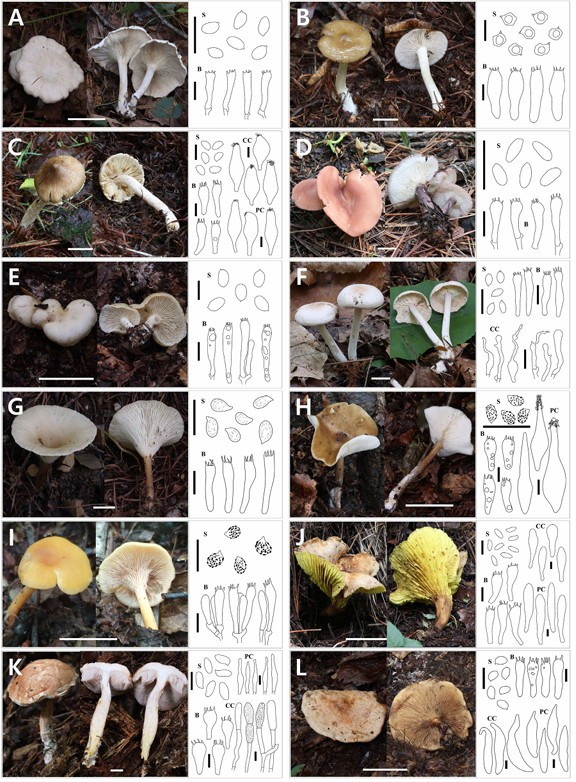
Fig. 1. Basidiocarps and microscopic structures of 12 fungal species: (A) Clitocybe tomentosa, (B) Entoloma lupinum, (C) Inocybe virgatula, (D) Calocybe vinacea, (E) Lyophyllum turcicum, (F) Gymnopus sinopolyphyllus, (G) Infundibulicybe trachyspora, (H) Melanoleuca leucopoda, (I) Notholepista fistulosa, (J) Phylloporus biyangensis, (K) Veloporphyrellus gracilioides, and (L) Paxillus cuprinus. Scale bars: basidiocarps = 5 cm (A, E, H, J, L) and 1 cm (B, C, D, F, G, I, K); microscopic drawings = 10 μm (S, basidiospores; B, basidia; CC, cheilocystidia; PC, pleurocystidia).
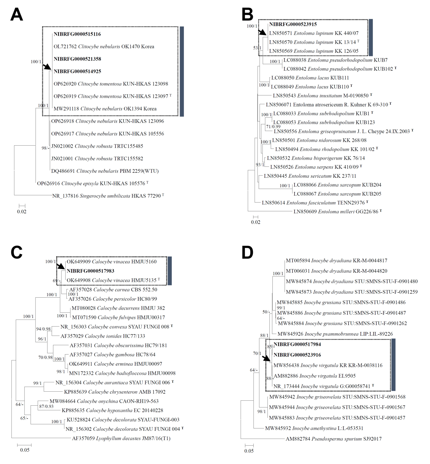
Fig. 2. Maximum likelihood phylogenetic trees of 12 macrofungal species newly recorded in Korea based on internal transcribed spacer (ITS) and large subunit ribosomal DNA (LSU) sequences. (A–J and L) Phylogenetic relationships of Clitocybe tomentosa, Entoloma lupinum, Inocybe virgatula, Calocybe vinacea, Lyophyllum turcicum, Gymnopus sinopolyphyllus, Infundibulicybe trachyspora, Melanoleuca leucopoda, Notholepista fistulosa, Phylloporus biyangensis, and Paxillus cuprinus inferred from ITS sequences. (K) Phylogenetic position of Veloporphyrellus gracilioides inferred from LSU sequences. Numbers above or below the branches indicate maximum likelihood bootstrap support values (MLBS ≥ 50%; left) and Bayesian posterior probabilities (BPP ≥ 0.90; right), respectively. Newly generated sequences are shown in bold. “T” indicates sequences from type specimens. Dotted boxes indicate clades containing the newly recorded Korean taxa, and arrows indicate the corresponding nodal support values. Vertical colored bars on the right indicate species boundaries. Double slashes (//) on branches indicate artificially shortened branch lengths.
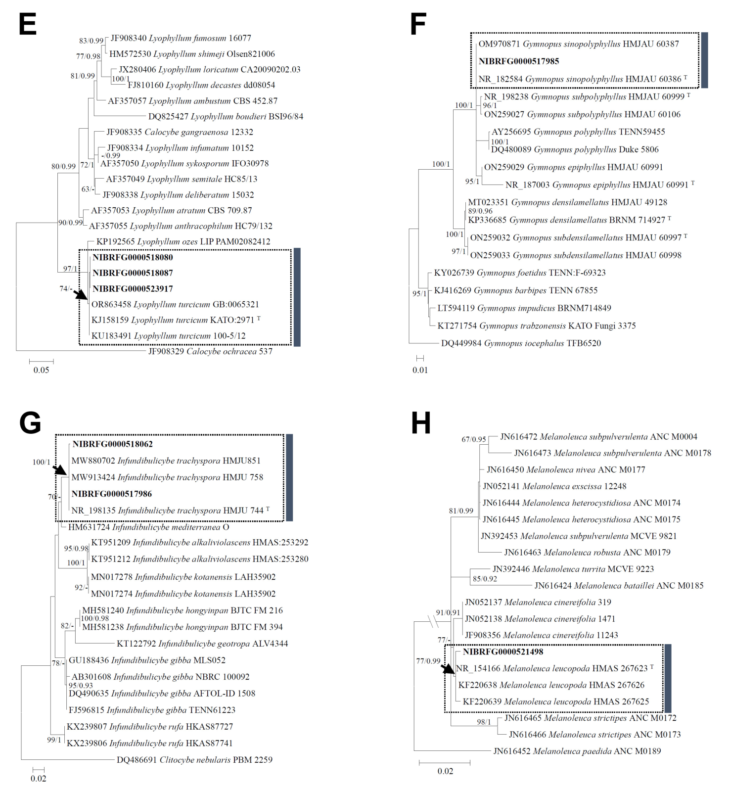
Fig. 2. Maximum likelihood phylogenetic trees of 12 macrofungal species newly recorded in Korea based on internal transcribed spacer (ITS) and large subunit ribosomal DNA (LSU) sequences. (A–J and L) Phylogenetic relationships of Clitocybe tomentosa, Entoloma lupinum, Inocybe virgatula, Calocybe vinacea, Lyophyllum turcicum, Gymnopus sinopolyphyllus, Infundibulicybe trachyspora, Melanoleuca leucopoda, Notholepista fistulosa, Phylloporus biyangensis, and Paxillus cuprinus inferred from ITS sequences. (K) Phylogenetic position of Veloporphyrellus gracilioides inferred from LSU sequences. Numbers above or below the branches indicate maximum likelihood bootstrap support values (MLBS ≥ 50%; left) and Bayesian posterior probabilities (BPP ≥ 0.90; right), respectively. Newly generated sequences are shown in bold. “T” indicates sequences from type specimens. Dotted boxes indicate clades containing the newly recorded Korean taxa, and arrows indicate the corresponding nodal support values. Vertical colored bars on the right indicate species boundaries. Double slashes (//) on branches indicate artificially shortened branch lengths. (continued)
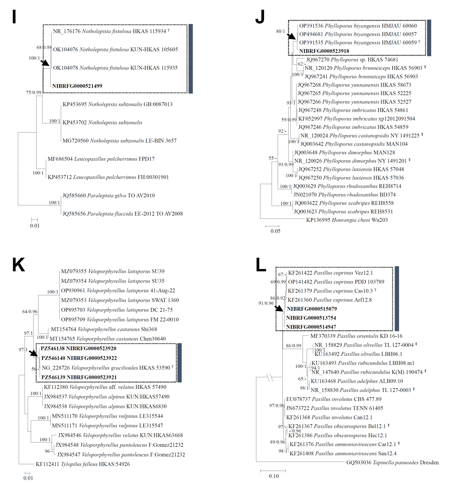
Fig. 2. Maximum likelihood phylogenetic trees of 12 macrofungal species newly recorded in Korea based on internal transcribed spacer (ITS) and large subunit ribosomal DNA (LSU) sequences. (A–J and L) Phylogenetic relationships of Clitocybe tomentosa, Entoloma lupinum, Inocybe virgatula, Calocybe vinacea, Lyophyllum turcicum, Gymnopus sinopolyphyllus, Infundibulicybe trachyspora, Melanoleuca leucopoda, Notholepista fistulosa, Phylloporus biyangensis, and Paxillus cuprinus inferred from ITS sequences. (K) Phylogenetic position of Veloporphyrellus gracilioides inferred from LSU sequences. Numbers above or below the branches indicate maximum likelihood bootstrap support values (MLBS ≥ 50%; left) and Bayesian posterior probabilities (BPP ≥ 0.90; right), respectively. Newly generated sequences are shown in bold. “T” indicates sequences from type specimens. Dotted boxes indicate clades containing the newly recorded Korean taxa, and arrows indicate the corresponding nodal support values. Vertical colored bars on the right indicate species boundaries. Double slashes (//) on branches indicate artificially shortened branch lengths. (continued)
(BS = 100%, PP = 1.00). Specimen NIBRFG0000521498 grouped with Melanoleuca leucopoda (BS = 77%, PP = 0.99), whereas specimen NIBRFG0000521499 clustered with Notholepista fistulosa (BS = 75%, PP = 0.99). Specimen NIBRFG0000523918 clustered with Phylloporus biyangensis (BS = 80%, PP = 1.00). Specimens NIBRFG0000515079, NIBRFG0000513754, and NIBRFG0000514947 belonged to the Paxillus cuprinus clade (BS = 91%, PP = 0.96). The identification of Veloporphyrellus gracilioides was supported by morphological characteristics and a phylogenetic analysis based solely on LSU rDNA sequences. LSU rDNA was used as the sole molecular marker because authenticated LSU reference sequences, including a sequence derived from the type material of V. gracilioides, were available for comparison. The Korean specimens NIBRFG0000523920, NIBRFG0000523922, and NIBRFG0000523921 formed a well-supported monophyletic clade with a type-derived sequence of V. gracilioides (BS = 97%, PP = 1.00), which supported their identification as V. gracilioides.
Detailed morphological descriptions and specimen data for each of the 12 newly recorded species are provided below.
Agaricales Underw. 1899
Clitocybaceae Vizzini, Consiglio & M. Marchetti 2020
1. Clitocybe tomentosa Z.M. He & Zhu L. Yang, in He, Chen, Bau, Wang, & Yang, Fungal Divers 123:14 (2023)
Korean name: Teol-kkal-ttae-gi-beo-seot (털깔때기버섯)
The Korean name refers to the densely tomentose surface of the pileus.
Pileus 40–100 mm in diameter, initially convex with a slightly inrolled margin, becoming plano-convex, then plane to slightly depressed at the center, and finally shallowly depressed to infundibuliform at maturity; surface densely tomentose to finely velutinous, with faint radial fibrils, whitish to pale cream, sometimes yellowish-brown and occasionally slightly darker at the center; margin incurved when young, later straight to undulate, with a tomentose to squamulose surface; context concolorous with pileus surface. Lamellae strongly decurrent, crowded to subcrowded, narrow to moderately broad, whitish to cream; lamellulae present; edges slightly irregular. Stipe 35–90 × 8–20 mm, central, cylindrical to slightly tapered downward, hollow; surface longitudinally fibrillose to slightly tomentose, white to pale cream; context concolorous with stipe surface.
Basidiospores (4–) 4.4–5.5 (–6) × 2.6–3.5 μm, Q = 1.42–1.89, Qm = 1.65 ± 0.17, ellipsoid to broadly ellipsoid, smooth, thin-walled, hyaline. Basidia 25–30.5 × 5–5.8 μm, clavate to cylindro-clavate, 4-spored, thin-walled, hyaline. Cheilocystidia and pleurocystidia absent.
Habitat: Solitary to scattered in deciduous forests, including those dominated by Quercus mongolica.
Specimens examined: KOREA, Gyeongsangbuk-do, Bonghwa-gun, Seokpo-myeon, Daehyeon-ri, solitary in deciduous forest, 37°04'09.2"N, 128°57'53.9"E, elevation 706 m, 28 Jun. 2023, Coll. JY Park and DG Choi, specimen no. NIBRFG0000514925 (QM20230628-15; GenBank accession No. PZ564178); Gyeongsangbuk-do, Bonghwa-gun, Seokpo-myeon, Daehyeon-ri, solitary in deciduous forest, 37°04'17.1"N, 128°58'04.8"E, elevation 698 m, 25 Jul. 2023, Coll. JY Park and DG Choi, specimen no. NIBRFG0000515116 (QM20230725-54; GenBank accession No. PZ564179); Gangwon-do, Jeongseon-gun, Gohan-eup, Gohan-ri, scattered in a deciduous forest dominated by Quercus mongolica, 37°12'16.3"N, 128°54'55.0"E, elevation 1,281 m, 25 Aug. 2025, Coll. JY Park and DG Choi, specimen no. NIBRFG0000521358 (QM20250825-08; GenBank accession No. PZ564180).
Remark: Clitocybe tomentosa is readily distinguished by its whitish to pale cream basidiomata, dense tomentose pileus surface, and strong decurrent lamellae. Morphologically, it closely resembles Clitocybe nebularis, which is widely reported in temperate regions. However, C. nebularis differs in having a smoother to finely fibrillose pileus surface and a distinct two-layered pileipellis composed of an irregular suprapellis and a regular subpellis [11]. The Korean specimens agree well with the original description of C. tomentosa in both macroscopic and microscopic features. Furthermore, several Korean collections previously identified as C. nebularis shared identical ITS sequences (MW291118 and OL721762) with those of C. tomentosa. Therefore, some historical Korean records of C. nebularis may represent misidentified specimens of C. tomentosa (Fig. 2A). Additional molecular and morphological studies are required to clarify the distribution and taxonomic status of these two species in Korea.
Entolomataceae Kotl. & Pouzar 1972
2. Entoloma lupinum Kokkonen, Mycol Progr 14(no. 116): 19 (2015)
Korean name: Neuk-dae-oe-dae-beo-seot (늑대외대버섯)
The Korean name was proposed based on the etymology of the species epithet and reflects the wolf-like, grayish-brown coloration of the basidiomata.
Pileus 20–50 mm in diameter, initially convex, later plano-convex to shallowly depressed at the center, with a low, broad umbo; surface distinctly viscid to glutinous when moist, smooth, olive-brown to yellowish-brown, slightly darker at the disk; margin translucently striated, especially when wet, slightly incurved when young, later straight to weakly undulate; context grayish-brown. Lamellae emarginate to adnate, moderately distant, pale cream to pale grayish brown when young, becoming pale pinkish as the spores mature; lamellulae present; edges even. Stipe 40–80 × 5–9 mm, central, cylindrical, slender, white to pale cream, smooth to finely fibrillose, slightly translucent toward the apex; base somewhat enlarged, covered with white tomentose mycelium; context concolorous with stipe surface.
Basidiospores (7.3–) 7.9–9.0 (–9.5) × (5.7–) 6.5–7.7 (–7.9) μm, Q = 1.09–1.30, Qm = 1.20 ± 0.08, subisodiametric, six-angled, smooth, thin-walled, hyaline. Basidia (32–) 34–39 × (8.5–) 9.3–11.7 (–12.4) μm, clavate to narrowly clavate, 4-spored, thin-walled, hyaline. Cheilocystidia 35.0–50.3 × 6.0–6.5 μm, cylindrical, flexuous, or clavate. Pleurocystidia absent.
Habitat: Solitary on soil in forests dominated by Larix kaempferi.
Specimen examined: KOREA, Gangwon-do, Taebaek-si, Changjuk-dong, solitary on soil in Larix kaempferi forest, 37°13'50.7"N, 128°55'40.9"E, elevation 931 m, 24 Jul. 2023, Coll. JY Park and DG Choi, specimen no. NIBRFG0000523915 (QM20230724-17; GenBank accession No. PZ564181).
Remark: Entoloma lupinum is characterized by a distinctly viscid, olive-brown pileus with a translucently striated margin, moderately distant, pale lamellae, and a slender, whitish stipe with a basal tomentum. Entoloma lupinum resembles E. radicipes but differs in having less fragile basidioma and smaller spores. It also differs from E. griseopruinatum in having a less fibrillose stipe, smaller spores, and incrusted pileipellis hyphae [12].
Inocybaceae Jülich 1982
3. Inocybe virgatula Kühner, Bull Soc Nat Oyonnax 9(Suppl., Mém. hors sér. 1): 7 (1955)
Korean name: Mu-nui-ttam-beo-seot (무늬땀버섯)
The Korean name refers to the pattern formed by the longitudinal fibrils on the pileus.
Pileus 15–35 mm in diameter, initially conical to campanulate, later convex with a persistent, obtuse umbo; surface dry, distinctly radially fibrillose to virgate, especially toward the margin, and finely squamulose near the disk; color dark brown to yellowish brown, with a paler brown to ochraceous-brown margin that often splits with age; veil remnants occasionally present along the pileus margin; context whitish. Lamellae adnexed to narrowly adnate, moderately crowded, ventricose, pale cream to light beige, becoming slightly darker with maturity; edges whitish and uneven. Stipe 25–60 × 2–6 mm, central, cylindrical, slender, sometimes slightly enlarged toward the base; surface pale cream to light brownish, finely fibrillose to pruinose, especially near the apex; basal mycelium white; context whitish.
Basidiospores (7.7–) 8–9.5 (–10) × (4.4–) 4.7–5.4 (–5.6) μm, Q = 1.56–1.89, Qm = 1.72 ± 0.10, ellipsoid to amygdaliform, smooth, thin-walled, pale brown. Basidia (23.5–) 27.8–36.5 (–40.0) × (7.2–) 8.0–10.7 (–11) μm, clavate, 4-spored, thin-walled, hyaline. Cheilocystidia 51–70.7 × 17.0–24.5 μm, mostly ventricose to broadly ventricose or lageniform to fusiform-lageniform, often with crystalline encrustations at the apex, thick-walled. Pleurocystidia 55.5–75 × 15.2–24 μm, similar to cheilocystidia, ventricose, lageniform to utriform-lageniform, often with crystalline encrustations at the apex, thick-walled.
Habitat: Solitary on soil in forests dominated by Larix kaempferi.
Specimens examined: KOREA, Gangwon-do, Taebaek-si, Changjuk-dong, solitary on soil in Larix kaempferi forest, 37°13'51.4"N, 128°56'03.7"E, elevation 907 m, 24 Jul. 2023, Coll. JY Park and DG Choi, specimen no. NIBRFG0000517984 (QM20230724-06; GenBank accession No. PZ564182); Gangwon-do, Jeongseon-gun, Gohan-eup, Gohan-ri, solitary on soil in Larix kaempferi forest, 37°08'22.2"N, 128°53'59.7"E, elevation 1,210 m, 23 Sep. 2025, Coll. JY Park and DG Choi, specimen no. NIBRFG0000523916 (QM20250923-17; GenBank accession No. PZ564183).
Remark: Inocybe virgatula is characterized by small- to medium-sized basidioma with a brown to dark-brown fibrillose pileus, usually with a distinct, low umbo; pale-cream to light-ochraceous lamellae; and a whitish to pale-brownish stipe. Morphologically, I. virgatula is easily confused with several smooth-spored brown species of Inocybe, particularly taxa within the so-called “fuscidula-complex” and allied groups. Among morphologically similar taxa, I. glabripes differs in having narrower cystidia and a more uniformly fibrillose pileus that lacks the distinctly virgate appearance characteristic of I. virgatula [13,14]. The Korean material agrees well with previous descriptions of the species in both macromorphological and micromorphological characteristics.
Lyophyllaceae Jülich 1982
4. Calocybe vinacea J.Z. Xu & Yu Li, in Xu, Yu, Suwannarach, Jiang, Zhao & Li, J. Fungi 7(12, no. 1101): 8 (2021)
Korean name: Po-do-saek-bam-beo-seot (포도색밤버섯)
The Korean name refers to the characteristic wine-red or grape-like coloration of the pileus.
Pileus 25–55 mm in diameter, convex to plano-convex when young, shallowly depressed with an obtuse umbo; surface smooth, dry to slightly moist, pastel pink, vinaceous-pink to dull reddish pink, darker toward the center; margin initially incurved, later uplifted, and irregularly lobed or wavy; context thin to moderately fleshy, whitish. Lamellae adnate to slightly subdecurrent, moderately crowded, white to cream; lamellulae present in several series; edges entire. Stipe 30–60 × 4–8 mm, central, cylindrical to slightly enlarged downward, longitudinally striated, finely fibrillose, pale violaceous- to grayish-vinaceous, darker in the lower portion; base white tomentose.
Basidiospores (4–) 4.3–5.8 (–6.3) × (2–) 2.2–2.5 (–2.8) μm, Q = 1.93–2.40, Qm = 2.14 ± 0.20, oblong to elongate-ellipsoid, smooth, thin-walled, hyaline. Basidia (18.5–) 19.0–23.5 (–23.9) × (4.6–) 4.9–6 μm, clavate, 4-spored, thin-walled, hyaline. Cheilocystidia and pleurocystidia absent.
Habitat: Solitary in Pinus densiflora-dominated forests.
Specimen examined: KOREA, Gangwon-do, Taebaek-si, Geumcheon-dong, solitary in Pinus densiflora-dominated forests, 37°05'17.1"N, 128°58'02.7"E, elevation 857 m, 27 Jun. 2023, Coll. JY Park and DG Choi, specimen no. NIBRFG0000517983 (QM20230627-02; GenBank accession No. PZ564184).
Remark: Calocybe vinacea is characterized by pastel-pink to vinaceous-pink basidiomata, adnate to slightly subdecurrent lamellae, and narrow, oblong basidiospores. It is similar to C. rubra but differs in having a paler vinaceous coloration, a more slender stipe, and narrower basidiospores [15]. The Korean collection differs slightly from the original description in having a somewhat stronger violaceous tint on the stipe. However, these differences appear to represent minor intraspecific variation that may be associated with ecological conditions or the developmental stage.
5. Lyophyllum turcicum Sesli, Vizzini & Contu, Turk J Bot 39:515 (2015)
Korean name: Dam-gal-man-ga-dak-beo-seot (담갈만가닥버섯)
The Korean name refers to the pale-brown to light-brown coloration of the basidiomata.
Pileus 18–52 mm in diameter, fleshy yet fragile, initially convex to broadly convex, becoming plano-convex to shallowly depressed at maturity; surface smooth, glabrous, moist, pale beige brown to grayish brown, and darker toward the center; margin incurved when young, later straight to weakly undulate, occasionally splitting at maturity; context white, up to 4 mm thick at the pileus center. Lamellae adnate to slightly decurrent, moderately crowded, whitish to pale cream, becoming faintly grayish cream with age; edges even. Stipe 28–65 × 4–11 mm, central, cylindrical to slightly clavate, sometimes curved or twisted, longitudinally fibrillose-pruinose, whitish to pale grayish brown; basal mycelium white and conspicuous.
Basidiospores 5–6 (–6.5) × (3.1–) 3.2–4.0 (–4.2) μm, Q = 1.43–1.69, Qm = 1.56 ± 0.12, ellipsoid to elongate-ellipsoid, smooth, usually containing oil droplets, thin-walled, hyaline. Basidia (26–) 26.4–37.4 (–39.5) × (5–) 5.5–7 μm, clavate, mostly 2–4-spored, with basal clamp connections, thin-walled, hyaline. Cheilocystidia and pleurocystidia absent. Marginal cells 18–38 × 2.8–4.5 μm, filiform-fusiform to cylindro-flexuose, thin-walled, hyaline, often irregularly curved.
Habitat: Gregarious on soil in forests dominated by Larix kaempferi or Quercus mongolica.
Specimens examined: KOREA, Gangwon-do, Taebaek-si, Changjuk-dong, gregarious on soil in Larix kaempferi forests, 37°13'51.2"N, 128°55'37.2"E, elevation 936 m, 26 Sep. 2024, Coll. JY Park and DG Choi, specimen no. NIBRFG0000523917 (QM20240926-49; GenBank accession no. PZ564185); Gangwon-do, Jeongseon-gun, Gohan-eup, Gohan-ri, gregarious in a deciduous forest dominated by Quercus mongolica, 37°12'12.7"N, 128°54'53.8"E, elevation 1,271 m, 27 Sep. 2024, Coll. JY Park and DG Choi, specimen no. NIBRFG0000518080 (QM20240927-01; GenBank accession no. PZ564186); Gangwon-do, Jeongseon-gun, Gohan-eup, Gohan-ri, gregarious in a deciduous forest dominated by Quercus mongolica, 37°12'14.2"N, 128°54'53.8"E, elevation 1,276 m, 27 Sep. 2024, Coll. JY Park and DG Choi, specimen no. NIBRFG0000518087 (QM20240927-20; GenBank accession No. PZ564187).
Remark: Lyophyllum turcicum is characterized by fragile pale brown basidiomata, adnate to slightly decurrent lamellae, and ellipsoid basidiospores. Morphologically, L. turcicum resembles L. fumosum, L. brunneum, and L. pseudoloricatum but differs in several important characteristics. Lyophyllum fumosum possesses darker gray-brown basidiomata and globose to subglobose spores, whereas L. turcicum has distinctly elongated ellipsoid spores [16]. L. brunneum differs in having stouter basidiomata, darker pileus coloration, and broader spores measuring 6–7 × 4.5–5 μm. L. pseudoloricatum is distinguished by its elastic-cartilaginous context, absence of marginal cells, and broader basidiospores 6–8 × 4.5–6 μm [17,18].
Omphalotaceae Bresinsky 1985
6. Gymnopus sinopolyphyllus J.P. Li, Chang Tian Li & Yu Li, in Li, Pan, Li, Deng, Wang, Zhang, Li & Li, J. Fungi 8(4, no. 398): 9 (2022)
Korean name: Hwang-hoe-saek-kkot-ae-gi-beo-seot (황회색꽃애기버섯)
The Korean name refers to the reddish-orange and yellowish-gray coloration of the pileus.
Pileus 12–28 mm in diameter, initially hemispherical to convex, becoming plano-convex with age; surface smooth, dry, pale cream to yellowish-gray, occasionally with a reddish-orange tinge and faint beige discoloration at the center; margin entire and hygrophanous but not striated; context thin, whitish. Lamellae free to adnate, crowded, cream to pale yellowish white, lamellulae present in several tiers. Stipe 25–55 × 2–5 mm, central, cylindrical, slightly enlarged toward the base, hollow, whitish to pale cream, with a reddish-brown tinge at the apex; surface smooth to finely fibrillose; basal mycelium white.
Basidiospores 4.8–6.4 (–6.7) × (2.5–) 2.7–3.5 (–3.7) μm, Q = 1.63–2.12, Qm = 1.81 ± 0.16, ellipsoid to oblong, smooth, thin-walled, hyaline. Basidia (22–) 24.5–33 (–36) × (4.5–) 5–6.5 (–7) μm, clavate, 4-spored, thin-walled, hyaline. Cheilocystidia 25.8–40.5 × 4.1–6.2 μm, narrowly to irregularly clavate, rarely lageniform to narrowly utriform, frequently flexuous or irregularly curved, sometimes with one or more projections or irregularly branched outgrowths, thin-walled, scattered along the lamellar edge. Pleurocystidia absent.
Habitat: Gregarious on leaf litter in deciduous forests.
Specimen examined: KOREA, Gyeongsangbuk-do, Bonghwa-gun, Seokpo-myeon, Daehyeon-ri, gregarious on leaf litter in deciduous forests, 37°04'13.7"N, 128°58'00.1"E, elevation 700 m, 28 Jun. 2023, Coll. JY Park and DG Choi, specimen no. NIBRFG0000517985 (QM20230628-10; GenBank accession No. PZ564188).
Remark: Gymnopus sinopolyphyllus is distinguished by its small whitish basidiomata, crowded lamellae, slender stipe with a conspicuous basal tomentum, and small ellipsoid basidiospores. The Korean collection agrees well with the original description of G. sinopolyphyllus in both macromorphological and micromorphological characteristics. In addition, G. sinopolyphyllus closely resembles G. cystidiosus in macromorphological features. However, the original description of G. cystidiosus reports the presence of pleurocystidia [19], whereas these structures were consistently absent in the Korean specimen. Because the Korean specimen closely conforms to the diagnostic features of G. sinopolyphyllus, it is assigned to that species. Although Gymnopus densilamellatus is morphologically similar to G. sinopolyphyllus, it can be distinguished by its incrusted pileipellis hyphae [20].
Omphalinaceae Vizzini, Consiglio & M. Marchetti 2020
Infundibulicybe Harmaja 2003
Korean name: O-mok-kkal-ttae-gi-beo-seot-sok (오목깔때기버섯속)
The Korean name refers to the distinctly funnel-shaped and depressed pileus characteristic of species in the genus.
7. Infundibulicybe trachyspora J.Z. Xu, J.C. Qin & Yu Li, in Xu, Zhao, Yu, Idrees, Li & Qin, Curr Microbiol 79(no. 130): 2 (2022)
Korean name: Geo-chin-po-ja-o-mok-kkal-ttae-gi-beo-seot (거친포자오목깔때기버섯)
The Korean name refers to the distinctly rough (ornamented) basidiospores.
Pileus 20–60 mm in diameter, initially convex with a shallow central depression, becoming plano-depressed to distinctly infundibuliform with age; surface dry, smooth to finely fibrillose, pale grayish beige to pale ochraceous-beige, and slightly darker and brownish-beige at the center; margin initially involute, often irregularly undulate, and faintly translucently striate near the edge when moist. Lamellae decurrent, moderately crowded, cream to yellowish-white or ivory; lamellulae of one to three lengths; frequently forked and intervenose near the stipe; edges even. Stipe 25–60 × 5–7 mm, central, cylindrical to slightly tapered downward; surface dry, longitudinally fibrillose, grayish brown to reddish brown, sometimes ochraceous beige to pale brownish; base densely covered with white tomentum and adherent mycelium.
Basidiospores 6.4–8.5 (–9) × 3.5–4.5 (–4.8) μm, Q = 1.67–1.97, Qm = 1.81 ± 0.12, lacrymoid to shortly ellipsoid, apiculate, rugulose, hyaline. Basidia (22–) 23.4–34 (–36) × (4.4–) 4.9–6 (–6.5) μm, subclavate to clavate, 4-spored, thin-walled, hyaline. Cheilocystidia and pleurocystidia absent.
Habitat: Solitary on soil in mixed coniferous–deciduous and Larix forests.
Specimens examined: KOREA, Gyeongsangbuk-do, Bonghwa-gun, Seokpo-myeon, Daehyeon-ri, solitary in mixed coniferous–deciduous forests, 37°03'50.9"N, 128°59'25.9"E, elevation 650 m, 25 Jul. 2023, Coll. JY Park and DG Choi, specimen no. NIBRFG0000517986 (QM20230725-39; GenBank accession No. PZ564189); Gangwon-do, Taebaek-si, Changjuk-dong, solitary on soil in Larix forests, 37°13'50.9"N, 128°55'53.1"E, elevation 928 m, 26 Sep. 2024, Coll. JY Park and DG Choi, specimen no. NIBRFG0000518062 (QM20240926-03; GenBank accession No. PZ564190).
Remark: Infundibulicybe trachyspora is readily recognized by its pale-beige to brownish, infundibuliform pileus; moderately crowded, narrow lamellae; brown to dark reddish-brown, longitudinally fibrillose-striated stipe; and rugulose basidiospores. It is morphologically similar to Ampulloclitocybe clavipes, which also has pale-beige to brownish infundibuliform basidiomata; crowded, decurrent lamellae; and rugulose basidiospores. However, A. clavipes is readily distinguished by its characteristic bulbous-clavate stipe base, whereas I. trachyspora has a cylindrical stipe that lacks basal swelling [21].
Melanoleucaceae Locq. ex Vizzini, Consiglio & P. Alvarado 2024
8. Melanoleuca leucopoda X.D. Yu, in Yu, Lv, Ma, Li, Lin & Zhang, Mycoscience 55(6): 458 (2014).
Korean name: Som-teol-dae-bae-kkob-beo-seot (솜털대배꼽버섯)
The Korean name is derived from the conspicuous cottony tomentum at the stipe base.
Pileus up to 70 mm in diameter, initially convex with an involute margin, later becoming plane with a distinctly depressed center; surface smooth to slightly uneven, somewhat viscid when wet, dull ochraceous-brown to clay-brown or yellowish-brown, and darker toward the disk; margin uplifted, somewhat undulating, occasionally lobed in mature basidiomata; context white. Lamellae adnate to shallowly decurrent, crowded, broad, cream white to dull white; lamellulae of two or four lengths; edges even. Stipe 100 × 12 mm, central, cylindrical to slightly tapered downward; surface pale brownish to cream-brown, longitudinally fibrillose; base covered with abundant white tomentum.
Basidiospores (7.7–) 7.8–9.5 (–10) × (4.5–) 4.7–6.2 (–6.5) μm, Q = 1.41–1.75, Qm = 1.62 ± 0.11, ellipsoid to oblong, ornamented mainly with scattered, rounded warts and occasional elongated warts. Basidia (27–) 28.2–35.5 (–37) × (8–) 8.2–10.5 (–11.5) μm, clavate, 4-spored, hyaline. Pleurocystidia 42.3–77 × 8.3–15.3 μm, fusiform to lageniform, frequently with encrusted crystals at the apex, thick-walled, hyaline. Cheilocystidia absent.
Habitat: Solitary in deciduous forests.
Specimen examined: KOREA, Gangwon-do, Taebaek-si, Changjuk-dong, solitary in deciduous forests, 37°13'50.0"N, 128°55'53.1"E, elevation 924 m, 26 Sep. 2024, Coll. JY Park and DG Choi, specimen no. NIBRFG0000521498 (QM20240926-26; GenBank accession no. PZ564191).
Remark: Melanoleuca leucopoda was originally described as a species characterized by small- to medium-sized basidioma with a pale-brown to pinkish cinnamon pileus; a whitish, fibrillose stipe; crowded, pale lamellae; amyloid basidiospores ornamented with elongated warts; and fusiform hymenial cystidia with apical crystalline encrustations [22]. The Korean specimen agrees closely with the original description in most diagnostic characteristics, particularly its elongated spore ornamentation, fusiform pleurocystidia, and whitish, fibrillose stipe. However, the Korean material has a larger basidioma and slightly smaller basidiospores than those described in the protologue. Nevertheless, because only a single Korean specimen was available for examination, these differences may represent individual or environmental variation rather than taxonomically significant divergence. Phylogenetic analysis based on the rDNA ITS region supported the identification of the Korean specimen as M. leucopoda. Additional collections and further studies on Korean specimens are necessary to assess the morphological variation and taxonomic stability of this species in East Asia [22].
Paralepistaceae Vizzini, Consiglio & P. Alvarado 2024
Notholepista Vizzini & Contu 2012
Korean name: Sa-ma-gwi-bang-mang-i-beo-seot-sok (사마귀방망이버섯속)
The Korean name refers to the distinctly verrucose (warted) basidiospores, which represent one of the diagnostic characteristics of the genus.
9. Notholepista fistulosa Z.M. He & Zhu L. Yang, Mycol Progr 21(2, no. 26): 9 (2022)
Korean name: Hwang-saek-sa-ma-gwi-bang-mang-i-beo-seot (황색사마귀방망이버섯)
The Korean name refers to the bright-yellow to orange-yellow pileus and distinctly verrucose basidiospores.
Pileus 10–25 mm in diameter, shallowly depressed to weakly umbilicate or infundibuliform in shape; surface smooth, dry, becoming moist to slightly lubricous when wet, bright-yellow to orange-yellow, disk slightly darker orange; margin initially incurved, later expanded and somewhat undulate, not striate; context thin and fragile, yellowish-white. Lamellae decurrent to deeply decurrent, moderately crowded, thick, sometimes forked, pale cream to pale-orange cream; lamellulae present in several series. Stipe 20–45 × 2–4 mm, central, cylindrical, hollow to fistulose; surface smooth, yellow-orange, slightly paler toward apex; basal mycelium white tomentose; context yellowish-white.
Basidiospores (4–) 4.3–6 (–6.4) × 3–4.5 μm, Q = 1.16–1.49, Qm = 1.33 ± 0.12, broadly ellipsoid to subglobose, distinctly verrucose, hyaline, thin-walled. Basidia (24.5–) 26.6–36 × (5–) 5.4–7 (–7.5) μm, subcylindrical, 4-spored, hyaline. Pleurocystidia and cheilocystidia absent.
Habitat: Solitary in Quercus mongolica-dominated deciduous forests.
Specimen examined: KOREA, Gangwon-do, Jeongseon-gun, Gohan-eup, Gohan-ri, solitary in a deciduous forest dominated by Quercus mongolica, 37°12'07.5"N, 128°54'45.2"E, elevation 1,270 m, 21 Aug. 2023, Coll. JY Park and DG Choi, specimen no. NIBRFG0000521499 (QM20230821-30; GenBank accession No. PZ564192).
Remark: Notholepista fistulosa is characterized by small, omphalinoid basidiomata; a bright-yellow to orange-yellow pileus; decurrent lamellae; a slender, fistulose stipe; and distinctly verrucose basidiospores. This species is morphologically similar to N. subzonalis but has a distinctly hollow stipe, whereas that of N. subzonalis is solid or stuffed [23].
Boletales E.-J. Gilbert 1931
Boletaceae Chevall. 1826
10. Phylloporus biyangensis Yang Wang, Bo Zhang & Yu Li, in Wang, Wang, Dai, Qi, Zhang, Liu, Hu, Zhang, Li & Zhang, Front Microbiol 13(no. 1052948): 7 (2023)
Korean name: Hwang-saek-min-geu-mul-beo-seot (황색민그물버섯)
The Korean name refers to the characteristic bright-yellow lamellae of the species.
Pileus 76–100 mm in diameter, initially plano-convex, applanate to shallowly depressed with age; surface dry, velvety to finely tomentose, brown to yellowish-brown or ochraceous-brown, occasionally with irregular cracks or patches exposing paler context; margin initially involute, later wavy and somewhat lobed; context pale yellow to cream, relatively thick in the center, unchanging when injured. Hymenophore lamellate, distinctly decurrent, moderately distant to subdistant, thick, blunt-edged, frequently forked and interconnected, bright sulfur-yellow to greenish-yellow, and slightly darker with age or after handling. Stipe 35–45 × 8–12 mm, central to slightly eccentric, cylindrical or slightly tapered downward, solid; surface pale yellow near the apex, becoming pale brownish to ochraceous toward the base, with ridges formed by the decurrent lamellae on the upper portion and covered with fine, yellow squamules; basal mycelium cream to light yellow; context pale yellow.
Basidiospores (8–) 8.4–10.8 (–11.6) × (3.5–) 3.6–4.4 (–4.7) μm, Q = 2.11–2.67, Qm = 2.34 ± 0.19, ellipsoid to elongate-ellipsoid, with a suprahilar depression in side view, thin-walled, brownish-yellow in 5% KOH. Basidia (31–) 32.6–41 (–42.5) × (7.5–) 8–9.7 (–10.5) μm, subcylindrical, 4-spored. Pleurocystidia 55.4–110.6 × 10.7–22 μm, abundant, subfusiform, narrowly clavate to lageniform, thin-walled, hyaline. Cheilocystidia 54.4–75.4 × 13.2–23.5 μm, clavate to sublageniform, thin-walled, hyaline.
Habitat: Solitary in Pinus densiflora forests.
Specimen examined: KOREA, Gyeongsangbuk-do, Bonghwa-gun, Seokpo-myeon, Daehyeon-ri, solitary in Pinus densiflora forests, 37°03'50.1"N, 128°59'24.0"E, elevation 649 m, 25 Jul. 2023, Coll. JY Park and DG Choi, specimen no. NIBRFG0000523918 (QM20230725-49; GenBank accession No. PZ564193).
Remark: Phylloporus biyangensis is characterized by a brown to yellowish-brown, dry, tomentose pileus; bright-yellow, strongly decurrent lamellae; elongated basidiospores; and abundant, clavate to lageniform pleurocystidia and cheilocystidia. Morphologically, P. biyangensis resembles Phylloporus luxiensis which also has bright-yellow lamellae and a brownish pileus. However, P. luxiensis differs in having a white context and larger basidiospores (10–12 × 4.5–5 μm) [24,25]. In addition, the pileus surface of P. luxiensis is usually smoother and less distinctly tomentose than that of P. biyangensis.
Veloporphyrellus L.D. Gómez & Singer 1984
Korean name: Pi-mak-geu-mul-beo-seot-sok (피막그물버섯속)
The Korean name refers to the veil-like structures observed in members of the genus and their affinities with poroid boletes.
11. Veloporphyrellus gracilioides Yan C. Li & Zhu L. Yang, in Wu, Li, Zhu, Zhao, Han, Cui, Li, Xu & Yang, Fungal Divers 81:165 (2016)
Korean name: Ga-neun-pi-mak-geu-mul-beo-seot (가는피막그물버섯)
The Korean name refers to the characteristically slender basidiomata and stipe of the species.
Pileus 20–45 mm in diameter, initially convex, hemispherical to applanate at maturity; surface dry, finely tomentose to subtomentose, brown to reddish-brown or cinnamon-brown, occasionally paler toward the margin; margin extended; context up to 6 mm thick at the center of the pileus, whitish to pale cream, with no color change upon exposure. Hymenophore poroid; adnate to slightly depressed around the stipe apex; pores angular to honeycomb-like, 1–3/mm; surface pale pinkish-gray to grayish cream or whitish, becoming pale brownish-pink to dingy pinkish-buff with age; tubes 3–7 mm long, concolorous with the hymenophoral surface, without color change after injury. Stipe 30–70 × 4–8 mm, central, slender, cylindrical to slightly curved, solid; surface dry, fibrillose, pale brown to reddish-brown, usually paler toward the apex and whitish near the base, occasionally covered with white powder elements, without color change when injured; basal mycelium white.
Basidiospores (12.4–) 12.6–14.3 (–15) × (4.8–) 5.3–6.8 μm, Q = 2.05–2.48, Qm = 2.26 ± 0.17, subfusiform and inequilateral in side view, with a slight suprahilar depression, smooth, slightly thick-walled, pale-yellowish to brownish-yellow in 5% KOH. Basidia (33–) 33.8–41.3 (–42.5) × (11–) 11.4–13.5 (–14) μm, clavate, mostly 4-spored, rarely 2-spored, hyaline. Pleurocystidia 45.6–57 × 5.7–9.7 μm, narrowly lageniform, fusiform to subfusiform, thin-walled, hyaline. Cheilocystidia 72.3–113.3 × 6.7–14.2 μm, lageniform to broadly clavate or irregularly ventricose, usually with 1–2 septa or secondary septa; terminal cells 25.5–46.5 μm long.
Habitat: Solitary in forests dominated by Pinus densiflora and in mixed coniferous–deciduous forests.
Specimens examined: KOREA, Gyeongsangbuk-do, Bonghwa-gun, Seokpo-myeon, Daehyeon-ri, solitary in Pinus densiflora forests, 37°03'50.3"N, 128°59'24.2"E, elevation 650 m, 25 Jul. 2023, Coll. JY Park and DG Choi, specimen no. NIBRFG0000523920 (QM20230725-48; GenBank accession No. PZ546138); Gyeongsangbuk-do, Bonghwa-gun, Seokpo-myeon, Daehyeon-ri, in mixed forests dominated by Pinus densiflora, 37°04'15.2"N, 128°58'00.1"E, elevation 703 m, 25 Jul. 2023, Coll. JY Park and DG Choi, specimen no. NIBRFG0000523921 (QM20230725-64; GenBank accession No. PZ546139); Gangwon-do, Taebaek-si, Geumcheon-dong, solitary in mixed coniferous–deciduous forests, 37°05'32.4"N, 128°57'50.4"E, elevation 944 m, 16 Sep. 2025, Coll. JY Park and DG Choi, specimen no. NIBRFG0000523922 (QM20250916-10; GenBank accession No. PZ546140).
Remark: Veloporphyrellus gracilioides is characterized by small and slender basidiomata; a dry, brown to reddish-brown, tomentose pileus; a pale pinkish-gray, poroid hymenophore; a slender, fibrillose stipe; relatively large, ellipsoid basidiospores; and remarkably elongated cheilocystidia. Morphologically, V. gracilioides resembles Austroboletus gracilis in having slender basidiomata and dry, brownish pileus. However, A. gracilis differs in having an apparently reticulate stipe and pitted-verrucose basidiospores [26].
Paxillaceae Lotsy 1907
12. Paxillus cuprinus Jargeat, Gryta, J.-P. Chaumeton & Vizzini, in Jargeat, Chaumeton, Navaud, Vizzini & Gryta, Fungal Biol 118(1): 26 (2014)
Korean name: Gu-rit-bit-u-dan-beo-seot (구릿빛우단버섯)
The Korean name refers to the coppery coloration of the pileus.
Pileus 25–100 mm in diameter, initially convex with an involute margin, later becoming plano-convex to depressed; surface glabrous when dry, slightly viscid to sticky when wet, finely tomentose to subtomentose, pale cream, buff, ochraceous-brown to coppery-brown, usually darker toward the center; margin persistently inrolled when young, later undulate and irregular; context pale cream to yellowish, turning reddish brown to dark brown when bruised. Lamellae deeply decurrent, crowded, frequently forked and anastomosing, initially cream to pale yellow, later ochraceous-brown to dark brown, rapidly discoloring when bruised; edges concolorous or slightly paler. Stipe 25–70 × 5–15 mm, mostly central, sometimes slightly eccentric, cylindrical or tapering downward, often curved; surface fibrillose to finely tomentose, pale cream near the apex, reddish-brown to dark-brown toward the base, especially in mature basidiomata; basal mycelium whitish.
Basidiospores (6.7–) 7.2–8.4 (–8.6) × (4.7–) 4.8–5.6 (–5.8) μm, Q = 1.37–1.64, Qm = 1.51 ± 0.09, ovoid to broadly ellipsoid or amygdaliform, smooth, thin-walled. Basidia (33.5–) 35–43.6 (–47.5) × (7–) 8–10.8 (–11.5) μm, cylindro-clavate, 4-spored, smooth, hyaline. Pleurocystidia 45.7–98.6 × 12–19 μm, ventricose with a long neck, broadly fusiform to lageniform or subcylindrical, often with a subcapitate apex, flexuous, thin-walled, with brown pigment in 5% KOH. Cheilocystidia 52.4–96.8 × 12.7–19.6 μm, lageniform to broadly fusiform or irregularly lageniform with a subcapitate apex, thin-walled, with a brown pigment in 5% KOH.
Habitat: Solitary in mixed coniferous–deciduous, deciduous, and oak forests.
Specimens examined: KOREA, Gangwon-do, Taebaek-si, Geumcheon-dong, solitary in mixed coniferous–deciduous forests, 37°05'14.3"N, 128°57'43.9"E, elevation 923 m, 31 Aug. 2022, Coll. JY Park and DG Choi, specimen no. NIBRFG0000513754 (QM20220831-70; GenBank accession No. PZ564196); Gyeongsangbuk-do, Bonghwa-gun, Seokpo-myeon, Daehyeon-ri, solitary in deciduous forests, 37°04'09.1"N, 128°58'47.4"E, elevation 650 m, 28 Jun. 2023, Coll. JY Park and DG Choi, specimen no. NIBRFG0000514947 (QM20230628-28; GenBank accession No. PZ564197); Gangwon-do, Taebaek-si, Wol-dong, solitary in oak forests, 37°07'28.0"N, 128°53'59.2"E, elevation 965 m, 25 Jul. 2023, Coll. JY Park and DG Choi, specimen no. NIBRFG0000515079 (QM20230725-19; GenBank accession No. PZ564198).
Remark: Paxillus cuprinus is characterized by pale-buff to coppery-brown basidiomata and strongly decurrent lamellae that rapidly turn dark brown when bruised. Morphologically, it closely resembles P. involutus, which has long been confused with this species. However, P. involutus typically has darker brown basidiomata and broader basidiospores but lacks the characteristic coppery coloration frequently observed in P. cuprinus. Molecular studies have also shown that P. cuprinus represents an independent lineage distinct from P. involutus [27].
This study documents 12 newly recorded macrofungal species in Korea and indicates that the distributions of many macrofungal species in East Asia remain poorly understood. Several of these species, including Calocybe vinacea, Clitocybe tomentosa, Gymnopus sinopolyphyllus, Infundibulicybe trachyspora, Notholepista fistulosa, and Phylloporus biyangensis, were described relatively recently, primarily from China [11,15,20,21,23–26,28]. Their occurrence in Korea suggests that several species previously known mainly from China may have broader distributions throughout the temperate and montane forests of East Asia. Although the Baekdudaegan Mountain Range may provide suitable habitats and potential dispersal corridors for these fungi, its role in their dispersal requires further investigation.
These findings also suggest that previous records of morphologically similar fungi in Korea should be re-evaluated. Notably, some Korean specimens identified as Clitocybe nebularis share ITS sequences identical or nearly identical to those of C. tomentosa. Therefore, some historical records of C. nebularis in Korea may represent C. tomentosa. Similar misidentifications may also exist among other groups of morphologically similar taxa. Reexamination of historical Korean specimens through combined morphological and molecular analysis may reveal overlooked species and clarify their distributions [11,28].
The results of the present study also highlight the taxonomic value of several microscopic characters. The distinctly verrucose basidiospores of Infundibulicybe trachyspora clearly distinguish this species from other members of the I. gibba complex [21,28]. Similarly, the identification of Inocybe virgatula was supported by the presence of characteristic metuloid cystidia and a longitudinally fibrillose pileus [28]. These results demonstrate that molecular and morphological evidence are complementary for the delineation of species boundaries and enhancement of taxonomic stability within Agaricomycetes. Furthermore, the 12 species belong to diverse ecological groups. Several species, including Gymnopus sinopolyphyllus, Clitocybe tomentosa, Entoloma lupinum, and Melanoleuca leucopoda, are putative saprotrophs associated with the decomposition of plant materials. In contrast, Paxillus cuprinus and Phylloporus biyangensis are presumed to be ectomycorrhizal based on their reported association with forest trees [25–27]. The occurrence of these ecologically diverse fungal groups in Taebaeksan National Park highlights the diversity of forest habitats in this region. The presence of fungi associated with both deciduous and coniferous trees further suggests that forest composition may influence fungal distribution. A limitation of this study is its reliance on a small number of DNA markers. Although ITS and selected LSU sequences provided useful evidence for species identification, analyses based on a single gene or a limited number of genes cannot fully resolve relationships among closely related species. Although ITS is widely used for fungal identification, it may have limited resolution among closely related taxa. Similarly, LSU is generally more suitable for resolving higher-level relationships than for distinguishing closely related species. Therefore, the phylogenetic results of this study should be interpreted in light of the limitations of available molecular data. Future research that incorporates multilocus or genome-scale data, additional specimens, and broader geographic sampling may provide further insights into species identification and evolutionary relationships.
In conclusion, these 12 newly recorded species enhance our understanding of Korean macrofungal diversity and the distribution of macrofungi in East Asia. Their occurrence in Korea suggests that some East Asian fungi may have broader distributions than previously recognized. However, additional sampling and molecular analyses are required to confirm their geographical ranges and ecological roles. Continued field surveys, reexamination of museum specimens, and multilocus phylogenetic analyses are essential to improve our understanding of macrofungal diversity and biogeography in Korea and throughout East Asia.
The following supplementary materials are available on the journal’s website: • Table S1. Characteristics of DNA datasets used for phylogenetic analyses • Table S2. MCMC convergence diagnostics for Bayesian analyses
No potential conflicts of interest relevant to this article were reported.
This study was supported by grants from the National Institute of Biological Resources (NIBR) funded by the Ministry of Climate, Energy, and Environment (MCEE) of the Republic of Korea (NIBR202502103-NIBR202602103).
1. Korea National Park Research Institute. Research of natural resources in Mt. Taebaeksan. Wonju: Korea National Park Research Institute; 2017. p. 893.
2. An JH, Park HJ, Lee SR, Seo IS, Nam GH, Kim JH. Analysis of environmental factors and change of vascular plant species along an elevational gradient in Baekdansa, Mt. Taebaeksan National Park. Korean J Environ Ecol 2019;33:378-401. https://doi.org/10.13047/KJEE.2019.33.4.378
[DOI]
3. Korea National Park Research Institute. Survey of park resources in Taebaeksan National Park. Wonju: Korea National Park Research Institute; 2023.
4. Park JY, Lee JS, Kim M, Lee H, Kim C, Kim NK. Newly recorded macrofungi from Taebaeksan National Park in Korea. Kor J Mycol 2023;51:313-34. https://doi.org/10.4489/KJM.20230033
[DOI]
5. Vilgalys R, Hester M. Rapid genetic identification and mapping of enzymatically amplified ribosomal DNA from several Cryptococcus species. J Bacteriol 1990;172:4238-46. https://doi.org/10.1128/jb.172.8.4238-4246.1990
[DOI][PubMed][PMC]
6. Gardes M, Bruns TD. ITS primers with enhanced specificity for basidiomycetes: Application to the identification of mycorrhizae and rust. Mol Ecol 1993;2:113-8. https://doi.org/10.1111/j.1365-294X.1993.tb00005.x
[DOI][PubMed]
7. White TJ, Bruns TD, Lee SB, Taylor JW. Amplification and direct sequencing of fungal ribosomal RNA genes for phylogenetics. In: Innis MA, Gelfand DH, Sninsky JJ, White TJ, editors. PCR protocols: A guide to methods and applications. San Diego: Academic Press; 1990. p. 315-22. https://doi.org/10.1016/B978-0-12-372180-8.50042-1
[DOI][PubMed][PMC]
8. Tamura K, Stecher G, Kumar S. MEGA11: Molecular evolutionary genetics analysis version 11. Mol Biol Evol 2021;38:3022-7. https://doi.org/10.1093/molbev/msab120
[DOI][PubMed]
9. Katoh K, Standley DM. MAFFT multiple sequence alignment software, version 7: Improvements in performance and usability. Mol Biol Evol 2013;30:772-80. https://doi.org/10.1093/molbev/mst010
[DOI][PubMed]
10. Ronquist F, Huelsenbeck JP. MrBayes 3: Bayesian phylogenetic inference under mixed models. Bioinformatics 2003;19:1572-4. https://doi.org/10.1093/bioinformatics/btg180
[DOI][PubMed]
11. He ZM, Chen ZH, Bau T, Wang GS, Yang ZL. Systematic arrangement within the family Clitocybaceae (Tricholomatineae, Agaricales): Phylogenetic and phylogenomic evidence, morphological data and muscarine-producing innovation. Fungal Divers 2023;123:1-47. https://doi.org/10.1007/s13225-023-00527-2
[DOI]
12. Kokkonen K. A survey of boreal Entoloma with emphasis on the subgenus Rhodopolia. Mycol Prog 2015;14:116. https://doi.org/10.1007/s11557-015-1135-y
[DOI]
13. Kuyper TW. A revision of the genus Inocybe in Europe. I. Subgenus Inosperma and the smooth-spored species of subgenus Inocybe. Persoonia Suppl 1986;3:1-247.
14. Bandini D, Oertel B, Eberhardt U. A fresh outlook on the smooth-spored species of Inocybe: Type studies and 18 new species. Mycol Prog 2021;20:1019-114. https://doi.org/10.1007/s11557-021-01712-w
[DOI]
15. Xu J, Yu X, Suwannarach N, Jiang Y, Zhao W, Li Y. Additions to Lyophyllaceae s.l. from China. J Fungi 2021;7:1101. https://doi.org/10.3390/jof7121101
[DOI][PubMed][PMC]
16. Consiglio G, Contu M. Il genere Lyophyllum P. Karst. emend. Kühner, in Italia. Riv Micol 2002;45:99-181.
17. Dähncke RM, Contu M, Vizzini A. New taxa in the genus Lyophyllum s.l. from La Palma (Canary Islands, Spain). Mycotaxon 2010;111:323-30. https://doi.org/10.5248/111.323
[DOI]
18. Sesli E, Vizzini A, Contu M. Lyophyllum turcicum (Agaricomycetes: Lyophyllaceae), a new species from Turkey. Turk J Bot 2015;39:512-9. https://doi.org/10.3906/bot-1407-16
[DOI]
19. Hu JJ, Song LR, Tuo YL, Zhao GP, Lei Y, Zhang B, Li Y. Multiple evidences reveal new species and a new record of smelly Gymnopus (Agaricales, Omphalotaceae) from China. Front Microbiol 2022;13:968617. https://doi.org/10.3389/fmicb.2022.968617
[DOI][PubMed][PMC]
20. Li JP, Pan MC, Li Y, Deng CY, Wang XM, Zhang BX, Li CT, Li Y. Morpho-molecular evidence reveals four novel species of Gymnopus (Agaricales, Omphalotaceae) from China. J Fungi 2022;8:398. https://doi.org/10.3390/jof8040398
[DOI][PubMed][PMC]
21. Xu J, Zhao W, Yu X, Idrees M, Li Y, Qin J. Infundibulicybe trachyspora, a new species from Northeastern China based on morphology and molecular phylogeny. Curr Microbiol 2022;79:130. https://doi.org/10.1007/s00284-022-02808-6
[DOI][PubMed]
22. Yu XD, Lv SX, Ma D, Li FF, Lin Y, Zhang L. Two new species of Melanoleuca (Agaricales, Basidiomycota) from northeastern China, supported by morphological and molecular data. Mycoscience 2014;55:456-61. https://doi.org/10.1016/j.myc.2014.01.007
[DOI]
23. He ZM, Yang ZL. The genera Bonomyces, Harmajaea and Notholepista from Northwestern China: Two new species and a new record. Mycol Prog 2022;21:26. https://doi.org/10.1007/s11557-022-01786-0
[DOI]
24. Zeng NK, Tang LP, Yang ZL. Type studies on two species of Phylloporus (Boletaceae, Boletales) described from southwestern China. Mycotaxon 2011;117:19-28. https://doi.org/10.5248/117.19
[DOI]
25. Wang Y, Wang LY, Dai D, Qi ZX, Zhang ZH, Liu YJ, Hu JJ, Zhang P, Li Y, Zhang B. Boletaceae in China: Taxonomy and phylogeny reveal a new genus, two new species, and a new record. Front Microbiol 2023;13:1052948. https://doi.org/10.3389/fmicb.2022.1052948
[DOI][PubMed][PMC]
26. Wu G, Li YC, Zhu XT, Zhao K, Han LH, Cui YY, Li F, Xu JP, Yang ZL. One hundred noteworthy boletes from China. Fungal Divers 2016;81:25-188. https://doi.org/10.1007/s13225-016-0375-8
[DOI]
27. Jargeat P, Chaumeton JP, Navaud O, Vizzini A, Gryta H. The Paxillus involutus (Boletales, Paxillaceae) complex in Europe: Genetic diversity and morphological description of the new species Paxillus cuprinus, typification of P. involutus s.s. and synthesis of species boundaries. Fungal Biol 2014;118:12-31. https://doi.org/10.1016/j.funbio.2013.10.008
[DOI][PubMed]
28. Vizzini A, Alvarado P, Consiglio G, Marchetti M, Xu J. Family matters inside the order Agaricales: Systematic reorganization and classification of incertae sedis clitocyboid, pleurotoid and tricholomatoid taxa based on an updated 6-gene phylogeny. Stud Mycol 2024;107:67-148. https://doi.org/10.3114/sim.2024.107.02
[DOI][PubMed][PMC]