
Yu-Na Choi, Ji-Won Kim, Seong-Eun Kim, and Ahn-Heum Eom*
Department of Biology Education, Korea National University of Education, Cheongju 28173, Korea
*Corresponding author: eomah@knue.ac.kr
Korean Journal of Mycology (Kor J Mycol) 2026 September, Volume 54, Issue 3, pages 275-285.
https://doi.org/10.4489/kjm.2026.54.3.5
Received on August 05, 2026, Revised on August 31, 2026, Accepted on September 04, 2026, Published on September 30, 2026.
© 2026 THE KOREAN SOCIETY OF MYCOLOGY.
This is an Open Access article distributed under the terms of the Creative Commons Attribution Non-Commercial License (http://creativecommons.org/licenses/by-nc/4.0/) which permits unrestricted non-commercial use, distribution, and reproduction in any medium, provided the original work is properly cited.
Biodegradable polyester, Fungal diversity, Marine macroalgae, Polycaprolactone, Seaweed endophytes
Plastic accumulation is a persistent environmental problem in terrestrial and marine ecosystems. Global plastic waste generation increased from 156 million tonnes (Mt) in 2000 to 353 Mt in 2019 [1,2], and substantial quantities of discarded plastics continue to enter natural habitats [3–6]. Although biodegradable polymers can reduce long-term persistence in some applications, their degradation depends strongly on the polymer properties, environmental conditions, and microorganisms and enzymes present at the disposal site [7,8].
Fungi belonging to genera such as Aspergillus, Penicillium, Trichoderma, and Fusarium have been screened for their abilities to alter or hydrolyze the ester bonds of synthetic polyesters such as polycaprolactone (PCL), a semi-crystalline aliphatic polymer [9,10]. However, most fungal screening studies have focused on terrestrial isolates, whereas the corresponding functional diversity of marine-derived fungi remains poorly characterized [11].
Marine macroalgae provide structurally and chemically distinct habitats for endophytic fungi, which produce extracellular enzymes such as esterases, lipases, and cutinase-like hydrolases to colonize the algal tissues [12–14]. As some of these enzymes act on ester-containing synthetic polymers, macroalgal endophytes are a relevant source of fungi for physiological screening.
The objectives of this study were to isolate and identify culturable endophytic fungi from the macroalgal species Ulva australis, Gracilaria vermiculophylla, and Sargassum thunbergii collected from the Dadohae coastal region of Korea; screen representative fungal isolates that possess PCL-hydrolyzing capability; and characterize the active strains using internal transcribed spacer (ITS) sequences and additional taxonomically informative loci. The plate-clearing assay was used in this study, and although it does not establish complete polymer biodegradation, it is useful for detecting extracellular hydrolytic activity and selecting strains for subsequent quantitative analyses.
Healthy asymptomatic specimens of Ulva australis (Chlorophyta), Gracilaria vermiculophylla (Rhodophyta), and Sargassum thunbergii (Ochrophyta) were collected from the intertidal zones of Aphaedo, Jaeundo, and Chupodo in Shinan-gun, Jeollanam-do, Republic of Korea, between June 17 and 18, 2025 (Fig. 1). The thalli were stored in sterile bags with ambient seawater and transported to the laboratory within 12 h at 4°C.
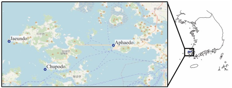
Fig. 1. Sampling sites of marine macroalgae in Shinan-gun, Korea.
The thalli were first surface sterilized to eliminate epiphytic microorganisms. The samples were washed under running tap water, cut into 1.0 × 0.5 cm segments, immersed in 70% ethanol for 15 s, and rinsed with sterile seawater. Then, the segments were dried on sterile tissue paper and plated onto dichloran rose bengal chloramphenicol agar (MBcell, Seoul, Korea) prepared with natural seawater and potato dextrose agar (PDA; Difco, Becton, Dickinson and Company, Franklin Lakes, NJ, USA) supplemented with chloramphenicol. The plates were incubated at 25°C in the dark. Emerging hyphae were subcultured on fresh PDA to obtain pure axenic cultures.
Genomic DNA was extracted from 14-day-old cultures grown on PDA, using the HiGene Genomic DNA Prep Kit (Biofact, Daejeon, Korea). For all isolates, the ITS region was amplified using ITS1F and ITS4 [15,16]. For isolates exhibiting PCL-hydrolyzing activity, additional loci were amplified using the following primers: ACT-512F/ACT-783R for actin (ACT) [17], EF1-728F/EF1-986R [17] or EF1-983F/ EF1-1567R for translation elongation factor 1-alpha (TEF1) [18], TUB2Fd/TUB4Rd for beta-tubulin (TUB2) [19], CMD5/CMD6 for calmodulin (CaM) [20], and LR0R/LR7 for large subunit 28S nuclear ribosomal DNA (LSU) [21]. The sequences generated in this study were deposited in the GenBank database under the accession numbers listed in Table 1.
Table 1. GenBank accession numbers currently available for PCL-hydrolyzing fungal isolates obtained in this study
| Strain | Identification | Genetic markers | GenBank accession numbers |
|---|---|---|---|
| KNUE25S357 | Alternaria alternata | ITS, LSU | PZ467978, PZ463299 |
| KNUE25S381 | Aureobasidium proteae | ITS, LSU | PZ463285, PZ463300 |
| KNUE25S347 | Phlebia acerina | ITS, LSU | PZ463280, PZ463284 |
| KNUE25S362 | Cladosporium anthropophilum | ITS, TEF1, ACT | PZ467979, PZ468812, PZ468816 |
| KNUE25S404 | Cladosporium halotolerans | ITS, TEF1, ACT | PZ467980, PZ468813, PZ468817 |
| KNUE25S425 | Cladosporium rectoides | ITS, TEF1, ACT | PZ467984, PZ468815, PZ468819 |
| KNUE25S421 | Cladosporium tenuissimum | ITS, TEF1, ACT | PZ467981, PZ468814, PZ468818 |
| KNUE25S422 | Diaporthe sp. (D. eres complex) | ITS, TUB2 | PZ467982, PZ468808 |
| KNUE25S444 | Paraphoma vinacea | ITS, TUB2 | PZ467986, PZ468810 |
| KNUE25S424 | Penicillium olsonii | ITS, TUB2 | PZ467983, PZ468809 |
| KNUE25S434 | Stemphylium vesicarium | ITS, CaM | PZ467985, PZ468811 |
ITS, internal transcribed spacer; LSU, large subunit 28S nuclear ribosomal DNA; TEF1, translation elongation factor 1-α; ACT, actin; TUB2, β-tubulin; CaM, calmodulin.
The consensus sequences were compared with sequences in the GenBank database using BLASTN. Maximum-likelihood phylogenetic analyses were performed using IQ-TREE v3.0.1 [22]. For multilocus datasets, the loci were concatenated and analyzed using the partition scheme specified for each dataset. The best-fit substitution model for each partition was selected using ModelFinder Plus [23], and branch support was estimated using 1,000 ultrafast bootstrap replicates [24].
The PCL-hydrolyzing activity was evaluated for one representative isolate per taxon using the emulsified agar plate-clearing assay [25]. The assay medium contained 1% (w/v) PCL diol (Mw ≈ 530; RHAWN, Shanghai, China) dissolved in acetone and incorporated into mineral-salts agar in 90 mm Petri dishes. A 6 mm mycelial plug was inoculated at the center of each dish, and the plates were incubated at 25°C for 7 days. The radial distance from the margin of the fungal colony to the outer boundary of the clear zone was measured using a digital caliper. To account for asymmetrical halo formation, the longest and shortest radial distances from the colony margin to the outer boundary of the clear zone were measured and averaged. All assays were performed in triplicate, and the results are presented as the means ± standard deviations.
In total, 43 endophytic fungal isolates were obtained from the three macroalgal species and assigned to 26 taxa on the basis of their colony characteristics and ITS sequence comparisons. These isolates were predominantly ascomycetous. Among the strains showing PCL-hydrolyzing activity, Phlebia acerina was the only basidiomycete. The sampling sites are shown in Fig. 1.
One representative strain of each taxon was screened on PCL-containing agar. Eleven of the 26 taxa examined (~42%) formed visible halo zones (Table 2). Active strains were recovered from all three macroalgal hosts. Ulva australis yielded the highest number of active taxa, although differences among hosts were not statistically tested because the study was designed as a culture-based survey rather than a standardized comparison of isolation frequencies.
Table 2. Algal host, isolation site, and PCL halo-zone size (mean ± SD; n = 3) of 11 active marine endophytic fungi
| Strain | Sampling site | Host macroalga | Identified taxon | Halo zone (Mean ± SD, mm) |
|---|---|---|---|---|
| KNUE25S362 | Jaeun-do | Ulva australis | C. anthropophilum | 13.50 ± 2.65 |
| KNUE25S381 | Aphae-do | Gracilaria vermiculophylla | A. proteae | 8.83 ± 2.75 |
| KNUE25S421 | Chupo-do | Sargassum thunbergii | C. tenuissimum | 8.83 ± 1.44 |
| KNUE25S357 | Aphae-do | Ulva australis | A. alternata | 6.33 ± 4.49 |
| KNUE25S434 | Chupo-do | Sargassum thunbergii | S. vesicarium | 4.67 ± 2.08 |
| KNUE25S425 | Chupo-do | Ulva australis | C. rectoides | 5.83 ± 3.34 |
| KNUE25S424 | Chupo-do | Ulva australis | P. olsonii | 3.00 ± 2.65 |
| KNUE25S404 | Chupo-do | Ulva australis | C. halotolerans | 3.67 ± 1.04 |
| KNUE25S444 | Jaeun-do | Sargassum thunbergii | P. vinacea | 1.67 ± 1.15 |
| KNUE25S347 | Aphae-do | Gracilaria vermiculophylla | P. acerina | 1.50 ± 0.87 |
| KNUE25S422 | Chupo-do | Sargassum thunbergii | Diaporthe sp. | 1.67 ± 0.58 |
The size of the halo zone differed among the fungal strains. Cladosporium anthropophilum KNUE25S362 produced the largest mean halo zone (13.50 ± 2.65 mm), whereas Aureobasidium proteae KNUE25S381 and Cladosporium tenuissimum KNUE25S421 produced mean halo zones of 8.83 mm. Alternaria alternata, Stemphylium vesicarium, and Cladosporium rectoides showed intermediate values, whereas Paraphoma vinacea, Phlebia acerina, and a Diaporthe sp. affiliated with the Diaporthe eres complex produced relatively small halo zones (Table 2). Representative culture plates are shown in Fig. 2.
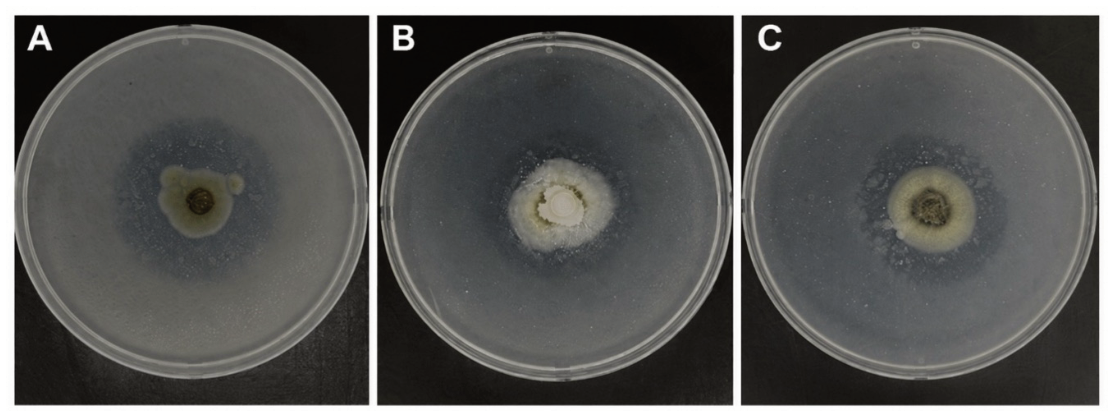
Fig. 2. Representative fungal strains exhibiting high polycaprolactone (PCL)-degrading activity on PCL-containing agar medium. A, Cladosporium anthropophilum KNUE25S362; B, Aureobasidium proteae KNUE25S381; C, Cladosporium tenuissimum KNUE25S421.
Four Cladosporium taxa produced halo zones, including the strain with the highest value. This is consistent with the broad ecological distribution and extracellular enzymatic capacity of Cladosporium species. A marine isolate of Cladosporium halotolerans was previously shown to degrade polyurethane [26], supporting the production of polyester-hydrolyzing enzymes by the genus Cladosporium. Aureobasidium species also produce extracellular esterases and lipases [27], which may explain the activity observed from Aureobasidium proteae. Considerable variation in PCL clear-zone formation has also been reported among fungi recovered from marine plastic debris [28]. However, because the size of the halo zone can be influenced by colony growth, enzyme secretion, diffusion through the agar, and substrate emulsification, the enzymatic activity observed in this study should be interpreted as a comparative screening response rather than a direct measure of the mass of polymer degraded [10,29].
The recovery of active strains from green, red, and brown macroalgae indicated that extracellular PCL-hydrolyzing activity was not restricted to a single host group in the present collection. Eleven of the 26 taxa examined produced clear zones, whereas a previous screening of 30 terrestrial fungal strains in Korea detected PCL- or polylactic acid-hydrolyzing activity in five strains [10]. These proportions should not be directly compared, because the fungal assemblages, substrates, and strain selection procedures differed between the two studies. Nevertheless, the findings from both studies, together with the results of a survey of marine plastic-associated fungi by Kim et al. [27], confirm that plate assays can be used to identify taxonomically diverse fungi with extracellular activity toward biodegradable polyesters. Confirmation of polymer biodegradation by the present isolates requires quantitative measurements such as polymer mass loss or molecular weight reduction, chemical analyses of the degradation products, and characterization of the responsible enzymes.
The ITS sequence-based phylogenetic relationships among representative isolates are shown in Fig. 3, and the multilocus trees used for the identification of selected strains are presented in Supplementary Fig. S1. Although the ITS sequences were sufficient for the preliminary placement of the isolates, the additional analyses of protein-coding loci improved the discrimination within species complexes and among closely related taxa. Multilocus sequence analysis is generally recommended for the accurate species delimitation of genera such as Diaporthe and Cladosporium, for which the ITS region alone often lacks sufficient resolution power [28–30].
Diaporthe strain KNUE25S422 shared 96.34% ITS and 97.66% TUB2 identities with the type strain within the Diaporthe eres complex. These values, together with the phylogenetic position of strain KNUE25S422, did not justify its identification at the species level. As mentioned above, species delimitation within Diaporthe, particularly the Diaporthe eres complex, generally requires the analyses of multiple protein-coding loci and morphological comparisons because the ITS sequence can show limited discriminatory power or intragenomic variation [29–31]. Therefore, because only the ITS and TUB2 data were available for strain KNUE25S422, it is conservatively reported as a Diaporthe sp. affiliated with the Diaporthe eres complex.
Strain KNUE25S444 was identified as Paraphoma vinacea on the basis of the ITS and TUB2 comparisons and its placement with reference strains in the multilocus analysis. This isolate was recovered from Sargassum thunbergii. Although this finding documents the occurrence of this strain among culturable fungi associated with the sampled macroalgal species, it does not establish host specificity or a new geographic record.
Within the genus Cladosporium, strains KNUE25S421 and KNUE25S362 grouped together with Cladosporium tenuissimum and Cladosporium anthropophilum, respectively. Although their ITS sequences closely matched those of the reference strains, the additional TEF1 and ACT sequence comparisons supported their phylogenetic placement. The use of ACT and TEF1 together with ITS for the species-level identification of Cladosporium is well established, whereas ITS alone often provides insufficient resolution among closely related species [28].
In conclusion, the three marine macroalgal species collected from the Dadohae coastal region harbored taxonomically diverse assemblages of culturable endophytic fungi. Eleven taxa produced clear zones on PCL-containing medium, with Cladosporium anthropophilum KNUE25S362 showing the strongest response. Multilocus analyses improved the identification of active strains and supported conservative identification of the divergent Diaporthe isolate. The strains identified in this study can be used as culture resources for subsequent quantitative studies of fungal enzymes and polyester biodegradation.
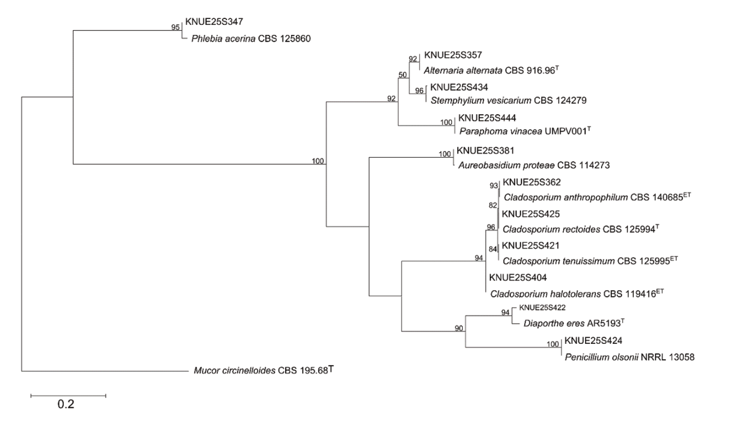
Fig. 3. ITS sequence-based maximum-likelihood phylogenetic tree of endophytic fungi isolated from marine macroalgae. Bootstrap values are shown at the nodes. Type (T) and ex-type (ET) strains are indicated. The scale bar represents nucleotide substitutions per site.
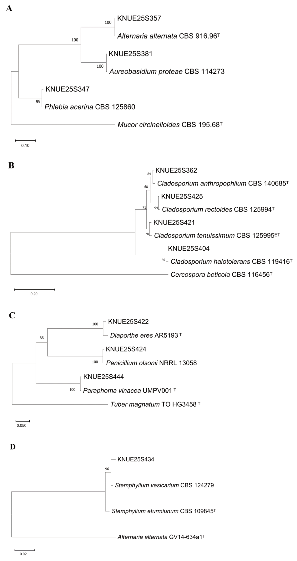
Fig. S1. Maximum-likelihood phylogenetic trees used to identify selected PCL-hydrolyzing fungal strains. Trees were inferred from (A) ITS and LSU, (B) ITS, TEF1, and ACT, (C) ITS and TUB2, and (D) ITS and CaM datasets. Bootstrap values are shown at the nodes. Type (T) and ex-type (ET) strains are indicated. Scale bars represent nucleotide substitutions per site. ITS, internal transcribed spacer; LSU, large subunit 28S nuclear ribosomal DNA; TEF1, translation elongation factor 1-α; ACT, actin; TUB2, β-tubulin; CaM, calmodulin.
The authors declare that they have no potential conflict of interest.
1. Pilapitiya PNT, Ratnayake AS. The world of plastic waste: A review. Clean Mater 2024;11:100220. https://doi.org/10.1016/j.clema.2024.100220
[DOI]
2. Vadera S, Khan S. A critical analysis of the rising global demand of plastics and its adverse impact on environmental sustainability. J Environ Pollut Manag 2021;3:105. https://doi.org/10.18875/2639-7269.3.105
[DOI]
3. Dokl M, Copot A, Krajnc D, Fan YV, Vujanović A, Aviso KB, Tan RR, Kravanja Z, Čuček L. Global projections of plastic use, end-of-life fate and potential changes in consumption, reduction, recycling and replacement with bioplastics to 2050. Sustain Prod Consum 2024;51:498-518. https://doi.org/10.1016/j.spc.2024.09.025
[DOI]
4. Geyer R, Jambeck JR, Law KL. Production, use, and fate of all plastics ever made. Sci Adv 2017;3:e1700782. https://doi.org/10.1126/sciadv.1700782
[DOI][PubMed][PMC]
5. Jambeck JR, Geyer R, Wilcox C, Siegler TR, Perryman M, Andrady A, Narayan R, Law KL. Plastic waste inputs from land into the ocean. Science 2015;347:768-71. https://doi.org/10.1126/science.1260352
[DOI][PubMed][PMC]
6. Lebreton LCM, van der Zwet J, Damsteeg JW, Slat B, Andrady A, Reisser J. River plastic emissions to the world’s oceans. Nat Commun 2017;8:15611. https://doi.org/10.1038/ncomms15611
[DOI][PubMed][PMC]
7. Bartnikowski M, Dargaville TR, Ivanovski S, Hutmacher DW. Degradation mechanisms of polycaprolactone in the context of chemistry, geometry and environment. Prog Polym Sci 2019;96:1-20. https://doi.org/10.1016/j.progpolymsci.2019.05.004
[DOI]
8. Kasuya K, Takagi K, Ishiwatari S, Yoshida Y, Doi Y. Biodegradabilities of various aliphatic polyesters in natural waters. Polym Degrad Stab 1998;59:327-32. https://doi.org/10.1016/S0141-3910(97)00155-9
[DOI]
9. Ekanayaka AH, Tibpromma S, Dai D, Xu R, Suwannarach N, Stephenson SL, Dao C, Karunarathna SC. A review of the fungi that degrade plastic. J Fungi 2022;8:772. https://doi.org/10.3390/jof8080772
[DOI][PubMed][PMC]
10. Lee SY, Ten LN, Das K, You YH, Jung HY. Biodegradative activities of fungal strains isolated from terrestrial environments in Korea. Mycobiology 2021;49:285-93. https://doi.org/10.1080/12298093.2021.1903131
[DOI][PubMed][PMC]
11. Zeghal E, Vaksmaa A, Vielfaure H, Boekhout T, Niemann H. The potential role of marine fungi in plastic degradation – a review. Front Mar Sci 2021;8:738877. https://doi.org/10.3389/fmars.2021.738877
[DOI]
12. Erbert C, Lopes AA, Yokoya NS, Furtado NAJC, Conti R, Pupo MT, Lopes JLC, Debonsi HM. Antibacterial compound from the endophytic fungus Phomopsis longicolla isolated from the tropical red seaweed Bostrychia radicans. Bot Mar 2012;55:435-40. https://doi.org/10.1515/bot-2011-0023
[DOI]
13. Murphy CA, Cameron JA, Huang SJ, Vinopal RT. Fusarium polycaprolactone depolymerase is cutinase. Appl Environ Microbiol 1996;62:456-60. https://doi.org/10.1128/aem.62.2.456-460.1996
[DOI][PubMed][PMC]
14. Tokiwa Y, Calabia BP, Ugwu CU, Aiba S. Biodegradability of plastics. Int J Mol Sci 2009;10:3722-42. https://doi.org/10.3390/ijms10093722
[DOI][PubMed][PMC]
15. Gardes M, Bruns TD. ITS primers with enhanced specificity for basidiomycetes – application to the identification of mycorrhizae and rusts. Mol Ecol 1993;2:113-8. https://doi.org/10.1111/j.1365-294X.1993.tb00005.x
[DOI][PubMed]
16. White TJ, Bruns TD, Lee SB, Taylor JW. Amplification and direct sequencing of fungal ribosomal RNA genes for phylogenetics. In: Innis MA, Gelfand DH, Sninsky JJ, White TJ, editors. PCR protocols: A guide to methods and applications. San Diego: Academic Press; 1990. p. 315-22. https://doi.org/10.1016/B978-0-12-372180-8.50042-1
[DOI][PubMed][PMC]
17. Carbone I, Kohn LM. A method for designing primer sets for speciation studies in filamentous ascomycetes. Mycologia 1999;91:553-6. https://doi.org/10.1080/00275514.1999.12061051
[DOI]
18. Rehner SA, Buckley E. A Beauveria phylogeny inferred from nuclear ITS and EF1-α sequences: Evidence for cryptic diversification and links to Cordyceps teleomorphs. Mycologia 2005;97:84-98. https://doi.org/10.1080/15572536.2006.11832842
[DOI][PubMed]
19. Woudenberg JHC, Aveskamp MM, de Gruyter J, Spiers AG, Crous PW. Multiple Didymella teleomorphs are linked to the Phoma clematidina morphotype. Persoonia 2009;22:56-62. https://doi.org/10.3767/003158509X427808
[DOI][PubMed][PMC]
20. Hong SB, Cho HS, Shin HD, Frisvad JC, Samson RA. Novel Neosartorya species isolated from soil in Korea. Int J Syst Evol Microbiol 2006;56:477-86. https://doi.org/10.1099/ijs.0.63980-0
[DOI][PubMed]
21. Vilgalys R, Hester M. Rapid genetic identification and mapping of enzymatically amplified ribosomal DNA from several Cryptococcus species. J Bacteriol 1990;172:4238-46. https://doi.org/10.1128/jb.172.8.4238-4246.1990
[DOI][PubMed][PMC]
22. Wong TKF, Ly-Trong N, Ren H, Demotte P, Baños H, Roger AJ, Susko E, Bielow C, De Maio N, Goldman N, et al. IQ-TREE 3: Phylogenomic inference software using complex evolutionary models. Mol Biol Evol 2026;43:msag117. https://doi.org/10.1093/molbev/msag117
[DOI][PubMed][PMC]
23. Kalyaanamoorthy S, Minh BQ, Wong TKF, von Haeseler A, Jermiin LS. ModelFinder: Fast model selection for accurate phylogenetic estimates. Nat Methods 2017;14:587-9. https://doi.org/10.1038/nmeth.4285
[DOI][PubMed]
24. Hoang DT, Chernomor O, von Haeseler A, Minh BQ, Vinh LS. UFBoot2: Improving the ultrafast bootstrap approximation. Mol Biol Evol 2018;35:518-22. https://doi.org/10.1093/molbev/msx281
[DOI][PubMed]
25. Zhang K, Hu J, Yang S, Xu W, Wang Z, Zhuang P, Grossart HP, Luo Z. Biodegradation of polyester polyurethane by the marine fungus Cladosporium halotolerans 6UPA1. J Hazard Mater 2022;437:129406. https://doi.org/10.1016/j.jhazmat.2022.129406
[DOI][PubMed]
26. Kudanga T, Mwenje E, Mandivenga F, Read JS. Esterases and putative lipases from tropical isolates of Aureobasidium pullulans. J Basic Microbiol 2007;47:138-47. https://doi.org/10.1002/jobm.200610207
[DOI][PubMed]
27. Kim SH, Lee JW, Kim JS, Lee W, Park MS, Lim YW. Plastic-inhabiting fungi in marine environments and PCL degradation activity. Antonie Van Leeuwenhoek 2022;115:1379-92. https://doi.org/10.1007/s10482-022-01782-0
[DOI][PubMed][PMC]
28. Bensch K, Braun U, Groenewald JZ, Crous PW. The genus Cladosporium. Stud Mycol 2012;72:1-401. https://doi.org/10.3114/sim0003
[DOI][PubMed][PMC]
29. Gomes RR, Glienke C, Videira SIR, Lombard L, Groenewald JZ, Crous PW. Diaporthe: A genus of endophytic, saprobic and plant pathogenic fungi. Persoonia 2013;31:1-41. https://doi.org/10.3767/003158513X666844
[DOI][PubMed][PMC]
30. Santos L, Alves A, Alves R. Evaluating multi-locus phylogenies for species boundaries determination in the genus Diaporthe. PeerJ 2017;5:e3120. https://doi.org/10.7717/peerj.3120
[DOI][PubMed][PMC]
31. Udayanga D, Castlebury LA, Rossman AY, Chukeatirote E, Hyde KD. Insights into the genus Diaporthe: Phylogenetic species delimitation in the D. eres species complex. Fungal Divers 2014;67:203-29. https://doi.org/10.1007/s13225-014-0297-2
[DOI]