
Seong-Eun Kim and Ahn-Heum Eom*
Department of Biology Education, Korea National University of Education, Cheongju 28173, Korea
*Corresponding author: eomah@knue.ac.kr
Korean Journal of Mycology (Kor J Mycol) 2026 September, Volume 54, Issue 3, pages 295-302.
https://doi.org/10.4489/kjm.2026.54.3.7
Received on August 20, 2026, Revised on September 22, 2026, Accepted on September 28, 2026, Published on September 30, 2026.
© 2026 THE KOREAN SOCIETY OF MYCOLOGY.
This is an Open Access article distributed under the terms of the Creative Commons Attribution Non-Commercial License (http://creativecommons.org/licenses/by-nc/4.0/) which permits unrestricted non-commercial use, distribution, and reproduction in any medium, provided the original work is properly cited.
Ectomycorrhiza, Pezizales, Sphaerosporella, Trichophaea, Truffle
Globose-spored, operculate discomycetes historically treated under the broad concept of Sphaerosporella brunnea have long presented taxonomic difficulties because of their overlapping morphological characteristics. Recent taxonomic studies incorporating morphological, ecological, and molecular data have clarified this complex [1,2]. True Trichophaea brunnea is now restricted to pyrophilous collections from burnt substrates, whereas Trichophaea scutelloides is recognized as a morphologically similar fungus occurring on unburned substrates [1].
S. brunnea and T. brunnea are combinations based on the same basionym, Peziza brunnea. Fungi identified as S. brunnea have frequently been reported in ectomycorrhizae of Tuber-inoculated nursery seedlings and are considered potential competitors of cultivated Tuber species [3–5]. Following the recent taxonomic revision of the T. brunnea complex, some collections from unburned nursery substrates have been referred to T. scutelloides [1]. To date, neither T. brunnea nor T. scutelloides has been reported in Korea.
During the cultivation of Tuber-inoculated hazelnut (Corylus sp.) seedlings in Korea, apothecia of a globose-spored discomycete appeared on the potting substrate. In this study, the morphological characteristics of the apothecia and the pure culture were examined, and phylogenetic analyses were conducted based on internal transcribed spacer (ITS) and large subunit (LSU) rDNA sequences to determine the identity of the fungus. Ectomycorrhizae formed on hazelnut roots were characterized and molecularly compared with apothecial and cultured materials. Based on morphological and phylogenetic evidence, T. scutelloides was reported for the first time in Korea, providing additional information on its ectomycorrhizal association with Corylus.
During the cultivation of Tuber-inoculated hazelnut (Corylus sp.) seedlings in a greenhouse at the Korea National University of Education (KNUE), Cheongju, Korea, apothecia appeared on a potting substrate consisting of vermiculite and perlite mixed at a 1:1 (v/v) ratio. Moreover, ectomycorrhizae morphologically distinct from those of Tuber were observed on the seedling roots. The seedlings, from which the apothecia and ectomycorrhizae examined in this study were obtained, had been sown in April 2022. They were initially maintained in a growth room at approximately 25°C, 60–70% relative humidity, and a 16-h light/8-h dark photoperiod, then transferred to a greenhouse. Apothecia and colonized root tips were collected from multiple pots in July 2026. A pure culture was obtained from fresh apothecial material. The resulting isolate was designated KNUE26T001 and maintained at KNUE.
Macroscopic characteristics of the apothecia were observed under a stereomicroscope (SZX7; Olympus, Tokyo, Japan). Hand sections and squash mounts were prepared from the hymenium, excipulum, and marginal tissues, and their microscopic characteristics were examined using a light microscope (Axio Imager A2; Carl Zeiss, Oberkochen, Germany). Measurements of ascospores (n = 50) and asci (n = 19) were taken from mature apothecia and reported as the (minimum–) mean (–maximum). For cultural characterization, the isolate was grown on malt extract agar (MEA; Kisan Bio, Seoul, Korea) and cornmeal agar (CMA; Kisan Bio, Seoul, Korea) at 25°C in darkness for 7 days. Colony growth, hyphal characteristics, and conidiogenous structures were examined. Ectomycorrhizae formed on Corylus roots were also characterized based on color, branching pattern, mantle surface, emanating hyphae, and rhizomorph formation.
Genomic DNA was extracted separately from the pure culture, fresh apothecium, and ectomycorrhizal root tips using a HiGene Genomic DNA Prep Kit (BioFact, Daejeon, Korea). The ITS region was amplified from all three materials using the primers ITS1F [6] and ITS4 [7] to confirm that the apothecium, pure culture, and ectomycorrhizae represented the same fungus. The LSU rDNA region was amplified from the pure culture and apothecium using the primers LR0R and LR5 [8]. The PCR products were sequenced by Macrogen (Sejong, Korea), and the resulting sequences were deposited in GenBank.
The newly generated ITS and LSU sequences were compared to sequences available in GenBank using BLASTn for preliminary identification. The reference sequences of T. scutelloides and closely related taxa were selected primarily from Van Vooren and Valade [1] (Table 1). Paranthracobia rehmii was used as the outgroup. Sequence alignment, concatenation, model selection, and phylogenetic analyses were performed using MEGA 12 [9]. ITS and LSU sequences were aligned separately and subsequently concatenated. The best-fit nucleotide substitution model was selected based on the Bayesian information criterion. Maximum likelihood analysis was performed using the Kimura two-parameter model with invariant sites (K2+I) [10] and 1,000 bootstrap replicates. Bootstrap values ≥70% are shown in the phylogenetic tree.
Table 1. Taxa and sequences used in the phylogenetic analysis
| Taxon / material | Voucher or isolate | ITS accession | LSU accession |
|---|---|---|---|
| Trichophaea scutelloides (culture) | KNUE26T001_C (this study) | PZ756363 | PZ756364 |
| Trichophaea scutelloides (apothecium) | KNUE26T001_A (this study) | PZ736826 | PZ736828 |
| Trichophaea scutelloides | RD 25.31.242.98 | MW476529 | MW546567 |
| Trichophaea scutelloides | LY NV 2022.11.02 | OR606626 | OR606632 |
| Trichophaea brunnea | LY NV 2021.06.03 | OR606625 | OR606631 |
| Trichophaea brunnea | Bru_JvP | MF066094 | MF066066 |
| Trichophaea ellipsoideospora | PC0714861 | MW476527 | MW546565 |
| Paranthracobia rehmii | PC0714858 | MW476530 | MW476531 |
ITS: internal transcribed spacer; LSU: large subunit.
The ITS sequences obtained directly from the apothecium and ectomycorrhizal root tips were identical to that obtained from the cultured isolate, confirming that all three materials represented the same fungus. In the maximum likelihood analysis of the concatenated ITS+LSU dataset, the Korean isolate clustered with the reference sequences of T. scutelloides and was clearly separated from T. brunnea and T. ellipsoideospora (Fig. 1).
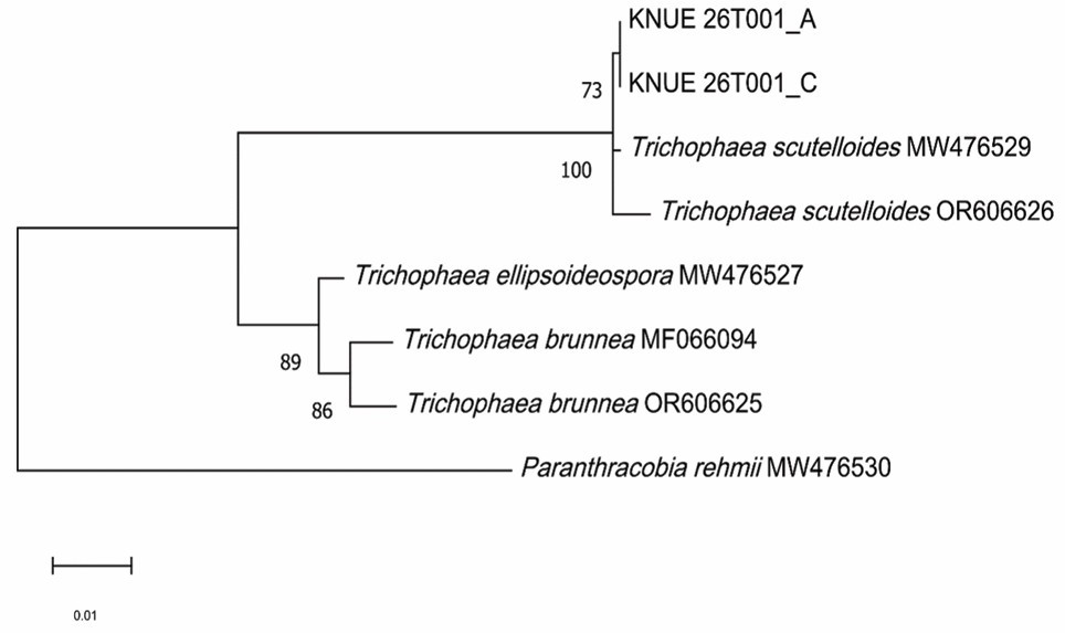
Fig. 1. Maximum likelihood phylogenetic tree based on the concatenated ITS and LSU rDNA dataset showing the placement of Trichophaea scutelloides KNUE26T001. The tree was constructed using the Kimura two-parameter model with invariant sites (K2+I). Bootstrap values ≥ 70% from 1,000 replicates are shown at the nodes. Paranthracobia rehmii was used as the outgroup. The scale bar represents 0.01 substitutions per site. ITS: internal transcribed spacer; LSU: large subunit.
Basionym: Peziza scutelloides Ellis, Bull. Torrey Bot. Club 9: 18 (1882).
Ascomata gregarious. Apothecia developing on the potting substrate of Tuber-inoculated seedlings, sessile, (3.07–)5.14(–10.26) mm in diameter (n = 8), discoid, often deformed by mutual pressure, orangish brown to fulvous brown. Hymenium orange-brown, smooth. External surface is orange and smooth. Margins fringed with dense brown hairs (Fig. 2A–E). Asci (87.64–)120.53(–155.87) × (12.17–)14.11(– 17.65) µm (n = 19), cylindrical, 8-spored, operculate, inamyloid, arising from croziers. Ascospores (10.62– )12.98(–14.38) µm in diameter (n = 50), globose, hyaline, smooth, uniguttulate with a single oil drop, thick-walled. Paraphyses cylindrical, apically widened, ± clavate to subcapitate, up to 10 µm wide, hyaline to diffusely orange, occasionally with light brown pigment deposited on the outer wall at the apex. Excipulum composed of textura angularis. Marginal hairs conical, tapering to acute apices, (63.44–)78.82(–89.56) × (4.04–)5.31(–7.98) µm. Excipular hairs were long, filamentous, and pale brown in color (Fig. 3A–F). Ectomycorrhizae (0.5–)0.95(–2.79) × (0.11–)0.18(–0.39) mm, brown, with a smooth mantle surface. Branching simple, mostly unbranched. Tips straight (linear, smooth-sided) and occasionally beaded (Fig. 2F). In culture, the hyphae on MEA were hyaline to pale brown and produced thick-walled brownish monilioid hyphae. Conidiophores and globose to subglobose conidiospores were observed on CMA and MEA (Fig. 4A–E).
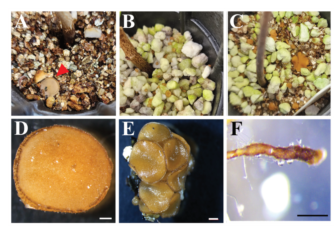
Fig. 2. Apothecia and ectomycorrhizae of Trichophaea scutelloides KNUE26T001 from Tuber-inoculated hazelnut seedlings in Korea. A–C, apothecia on potting substrate; the red arrowhead in A indicates an apothecium; D, single apothecium; E, gregarious apothecia; F, ectomycorrhizae on Corylus roots. Scale bars: D and E, 1 mm; F, 0.2 mm.
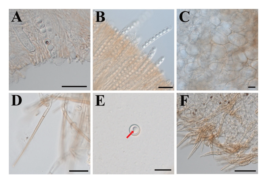
Fig. 3. Microscopic characteristics of the apothecia of Trichophaea scutelloides KNUE26T001. A, asci and paraphyses; B, asci; C, excipulum; D, excipular hairs; E, ascospore, with the red arrow indicating the single oil drop (guttule); F, marginal hairs. Scale bars: A, B, D, and F, 50 µm; C and E, 20 µm.
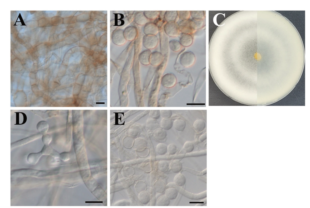
Fig. 4. Cultural characteristics of Trichophaea scutelloides KNUE26T001 on malt extract agar (MEA) and cornmeal agar (CMA). A, thick-walled monilioid hyphae on MEA; B, conidiophores and conidia on CMA; C, colony after 7 days on MEA; D, conidiophore with developing conidia on MEA; E, conidia on MEA. Scale bars: A, B, D, and E, 20 µm.
Note. The isolate examined in this study was identified as T. scutelloides based on its morphological characteristics and ITS+LSU phylogenetic analysis. Its occurrence on unburned substrates is consistent with the ecology of T. scutelloides. The morphological characteristics overlap considerably with those of T. brunnea, making it difficult to distinguish these taxa based on morphology alone (Table 2).
Table 2. Comparison of Trichophaea brunnea and T. scutelloides with the isolate examined in this study
| Character | T. brunnea [1] | T. scutelloides [1] | T. scutelloides (this study) |
|---|---|---|---|
| Substrate ecology | Burnt substrate; pyrophilous | Unburned substrate | Unburned nursery potting substrate |
| Apothecia | 1–5 mm; brown tones | 5–11 mm; fulvous to brown or orangish brown | (3.07–)5.14(–10.26) mm; orangish brown to fulvous brown |
| Ascospores | Globose, smooth, uniguttulate; 12–16.5 µm | Globose, smooth, uniguttulate; 13–15 µm | Globose, smooth, uniguttulate; (10.62–)12.98(–14.38) µm |
| Asci | 150–210 × 13–22 µm | (160–)210–270 × 12–16 µm | (87.64–)120.53(–155.87) × (12.17–)14.11(–17.65) µm |
| Ectomycorrhizae | Reported as ectomycorrhizal | Reported as ectomycorrhizal / associated with unburned substrates | (0.5–)0.95(–2.79) × (0.11–)0.18(– 0.39) mm, brown, smooth mantle, simple/mostly unbranched, observed on Corylus roots |
| Molecular placement | T. brunnea clade | T. scutelloides clade | T. scutelloides clade |
Specimen examined: KOREA. Cheongju, on potting substrate and roots of Tuber-inoculated Corylus sp. seedlings, July 2026, KNUE26T001 (NIBRFGC000515587); GenBank accession numbers: (ITS) PZ756363 (culture), PZ736826 (apothecium), PZ736827 (ectomycorrhiza), (LSU) PZ756364 (culture), and PZ736828 (apothecium).
This study provides the first record of T. scutelloides in Korea, supported by its morphological characteristics and ITS+LSU phylogenetic analysis. T. scutelloides is currently found in North America and Europe, with documented collections from the United States, Canada, France, and Germany [1]. Reference sequences used in the phylogenetic analysis were obtained from collections in France. The isolate from this study clustered with the reference sequences of T. scutelloides and was clearly separated from T. brunnea and T. ellipsoideospora. T. ellipsoideospora differs from T. scutelloides in having ellipsoid rather than globose ascospores and in its association with burnt substrates. The morphological characteristics of these species overlap considerably, which limits the reliability of morphology alone for species identification within this complex [1]. The shorter asci observed in the Korean material, compared with those reported by Van Vooren and Valade [1], may partly reflect variation in developmental stage. The occurrence of T. scutelloides on an unburned substrate in the present study is consistent with its known ecology, whereas true T. brunnea is regarded as a pyrophilous species associated with burnt substrates [1,2]. ITS sequences obtained from the apothecium, pure culture, and ectomycorrhizal root tips were identical, demonstrating that these materials represent the same fungus. This provides direct evidence that T. scutelloides forms ectomycorrhizae with Corylus under the cultivation conditions examined. Globose-spored fungi, previously identified as S. brunnea, have frequently been reported as ectomycorrhizal associates of Tuber-inoculated seedlings in European truffle nurseries [3–5], and some material from this ecological context is now referred to as T. scutelloides under the revised taxonomy [1]. Therefore, the present study extends the known geographic distribution of T. scutelloides to Korea and provides additional morphological and molecular documentation of its ectomycorrhizal association with Corylus.
No potential conflict of interest was reported by the authors.
This work was supported by a grant from the National Institute of Biological Resources (NIBR202602103), funded by the Ministry of Environment (MOE) of the Republic of Korea.
1. Van Vooren N, Valade F. Exploring the European Trichophaea-like discomycetes (Pezizales) using morphological, ecological and molecular data. Part 4: New insights in Trichophaea s. str. and Paranthracobia gen. nov. Ascomycete.org 2023;15:113-34. https://doi.org/10.25664/art-0376
[DOI]
2. Hughes KW, Case A, Matheny PB, Kivlin S, Petersen RH, Miller AN, Iturriaga T. Secret lifestyles of pyrophilous fungi in the genus Sphaerosporella. Am J Bot 2020;107:876-85. https://doi.org/10.1002/ajb2.1482
[DOI][PubMed][PMC]
3. Danielson RM. Ectomycorrhiza formation by the operculate discomycete Sphaerosporella brunnea (Pezizales). Mycologia 1984;76:454-61. https://doi.org/10.1080/00275514.1984.12023866
[DOI]
4. García-Montero LG, Galán R, Di Massimo G, Manjón JL, Díez J. Sphaerosporella brunnea (Alb. et Schwein.) Svrcek et Kubicka, un discomicete con incidencia en la truficultura e interes forestal. Rev Catalana Micol 1997;20:99-104.
5. Sánchez S, Gómez E, Martín M, De Miguel AM, Urban A, Barriuso J. Experiments on the life cycle and factors affecting reproduction of Sphaerosporella brunnea provide evidence for rapid asexual propagation by conidiospores and for homothallism in an ectomycorrhizal competitor of cultivated truffle species. Fungal Ecol 2014;8:59-65. https://doi.org/10.1016/j.funeco.2013.12.003
[DOI]
6. Gardes M, Bruns TD. ITS primers with enhanced specificity for basidiomycetes-application to the identification of mycorrhizae and rusts. Mol Ecol 1993;2:113-8. https://doi.org/10.1111/j.1365-294X.1993.tb00005.x
[DOI][PubMed]
7. White TJ, Bruns TD, Lee SB, Taylor JW. Amplification and direct sequencing of fungal ribosomal RNA genes for phylogenetics. In: Innis MA, Gelfand DH, Sninsky JJ, White TJ, editors. PCR protocols: A guide to methods and applications. San Diego: Academic Press; 1990. p. 315-22. https://doi.org/10.1016/B978-0-12-372180-8.50042-1
[DOI][PubMed][PMC]
8. Vilgalys R, Hester M. Rapid genetic identification and mapping of enzymatically amplified ribosomal DNA from several Cryptococcus species. J Bacteriol 1990;172:4238-46. https://doi.org/10.1128/jb.172.8.4238-4246.1990
[DOI][PubMed][PMC]
9. Kumar S, Stecher G, Suleski M, Sanderford M, Sharma S, Tamura K. MEGA12: Molecular Evolutionary Genetic Analysis version 12 for adaptive and green computing. Mol Biol Evol 2024;41:msae263. https://doi.org/10.1093/molbev/msae263
[DOI][PubMed][PMC]
10. Kimura M. A simple method for estimating evolutionary rates of base substitutions through comparative studies of nucleotide sequences. J Mol Evol 1980;16:111-20. https://doi.org/10.1007/BF01731581
[DOI][PubMed]