
Tae-Ho Kim, Yu-Na Choi, Seong-Eun Kim, Ji-Won Kim, and Ahn-Heum Eom*
Department of Biology Education, Korea National University of Education, Cheongju 28173, Korea
*Correspondence to eomah@knue.ac.kr
Korean Journal of Mycology (Kor J Mycol) 2026 June, Volume 54, Issue 2, pages 181-189.
https://doi.org/10.4489/kjm.2026.54.2.8
Received on March 26, 2026, Revised on June 08, 2026, Accepted on June 15, 2026, Published on June 30, 2026.
Copyright © The Korean Society of Mycology.
This is an Open Access article which is freely available under the Creative Commons Attribution-Non-Commercial 4.0 International License (CC BY-NC) (https://creativecommons.org/licenses/by-nc/4.0/).
Codium fragile, Diaporthe novem, Endophytic fungi, Green macroalga, Paradendryphiella arenariae
Endophytes are asymptomatic microbial symbionts that colonize the internal tissues of healthy plants, maintain intimate associations with their hosts, confer benefits under various developmental and environmental conditions, and may alter their lifestyles through dynamic interactions and co-evolution with the host plant [1]. In marine environments, endophytic fungi associated with macroalgae produce secondary metabolites that enhance host tolerance to environmental stress and provide defense against herbivores, thereby attracting increasing scientific interest [2]. Marine macroalgae-associated endophytes predominantly belong to the phylum Ascomycota, with genera such as Aspergillus, Penicillium, Fusarium, Alternaria, and Cladosporium being frequently reported in diverse seaweed hosts [2]. Despite their high ecological and biotechnological value, the biodiversity and functional roles of marine macroalgal endophytes remain underexplored on a global scale [3].
Codium fragile is a siphonous green macroalga widely distributed across the temperate rocky intertidal zones of the Pacific and Atlantic Oceans [4]. In undisturbed Korean coastal communities, C. fragile typically plays an understory role beneath dominant canopy-forming large brown algae, such as Sargassum spp. and Ecklonia cava [4]. In Korea, systematic investigations of the fungal diversity associated with marine macroalgae have only recently begun, focusing primarily on brown algae [2]. To our knowledge, the endophytic fungal communities associated with C. fragile in its native range in Korea have not been investigated.
A systematic survey was conducted with endophytic fungi isolated from healthy, asymptomatic tissues of C. fragile collected from the intertidal zones of Korea. Based on morphological characterization and multi-locus phylogenetic analyses, two endophytic fungal species were identified which were previously unrecorded in Korea: Paradendryphiella arenariae and Diaporthe novem. This study provides the first detailed morphological and molecular phylogenetic descriptions of these two newly recorded species and expands the current knowledge of marine algal-associated fungi in Korea.
Healthy, asymptomatic specimens of C. fragile without any visible wounds, necrotic lesions, or malformations were collected from the intertidal rocky shores of Uljin-gun (36°42′40.5″N, 129°28′31.7″E) and Pohang-si (35°59′44.8″N, 129°33′58.1″E), Gyeongsangbuk-do, Korea, in October 2025. The collected samples were placed in sterile plastic bags containing ambient seawater and were transported to the laboratory within 12 h under cool and shaded conditions. Upon arrival, specimens were immediately processed for surface sterilization and fungal isolation.
To eliminate epiphytic microbes and surface contaminants, algal thalli were thoroughly rinsed with running tap water and cut into approximately 1.0 cm segments [5]. Surface sterilization was performed by immersing the segments in 70% ethanol for 15 s, followed by rinsing thrice with sterile artificial seawater to remove any residual ethanol [6]. The sterilized segments were blotted dry using sterile cotton under aseptic conditions [7]. The dry segments were then placed on dichloran rose bengal chloramphenicol (Mbcell, Seoul, Korea) agar prepared with seawater and potato dextrose agar (PDA; Difco Lab., Detroit, USA) supplemented with seawater and chloramphenicol (100 µg/mL) to suppress bacterial growth [5]. The plates were incubated in the dark at 25°C to induce the growth of the endophytic fungi [8].
As fungal hyphae began to emerge from the internal tissues of C. fragile segments, they were transferred to fresh media to obtain pure isolates [8]. The pure strains were cultured on malt extract agar (MEA; Kisan Bio, Seoul, Korea) and PDA at 25°C in the dark to evaluate colony morphology, including growth rate, pigmentation, texture, and marginal features. Microscopic structures including conidiophores, conidiogenous cells, and conidia were observed and measured using a light microscope (Axio Imager A2; Carl Zeiss, Oberkochen, Germany) [9]. Spore dimensions were recorded from at least 20 random measurements to calculate average values and ranges.
Genomic DNA was extracted from fresh mycelia grown on PDA at 25°C using the HiGene Genomic DNA Prep Kit (Biofact, Daejeon, Korea) according to the manufacturer’s instructions. Polymerase chain reaction (PCR) was used to amplify the five molecular markers. The following primer sets were used for PCR amplification of fungal DNA regions: ITS1F/ITS4 for internal transcribed spacer (ITS) region [10,11], LR0R/LR5 for nuclear large subunit ribosomal DNA (LSU) [12], NS1/NS4 for nuclear small subunit ribosomal DNA (SSU) [13], EF1-728F/EF1-986R for translation elongation factor 1-alpha (tef1-α) [14], and RPB2-5f2/RPB2-7cr for RNA polymerase II second largest subunit (rpb2) [15]. PCR amplicons were verified by electrophoresis on a 1.5% agarose gel to confirm the expected sizes. DNA sequencing was performed by Macrogen Co., Ltd. (Sejong, Korea).
The obtained nucleotide sequences were compared with those in the GenBank database of the National Center for Biotechnology Information (NCBI) using the basic local alignment search tool (BLAST) to identify the closest related taxa and type strains. Sequence alignments were performed using MEGA11 software [16]. Concatenated datasets were prepared using appropriate markers for each genus. Phylogenetic trees were constructed using the maximum likelihood (ML) method under the best-fit nucleotide substitution model (K2+G), and branch robustness was evaluated using bootstrap analysis with 1,000 replicates. The isolated strains were deposited at the National Institute of Biological Resources (NIBR), Incheon, Korea, and their sequence data were registered in NCBI GenBank.
Paradendryphiella arenariae (Nicot) Woudenb. & Crous, Stud Mycol 75: 208 (2013) [MB#803752] Fig. 1
Morphological characteristics of KNUE25A105: On MEA, after 7 days of incubation at 25°C in the dark, the colony reached 35.0–41.0 mm in diameter. The colony was flat, circular, and exhibited a cottony texture with a pale yellow center and a gradually fading margin. The reverse side of the colony was lemon yellow in the center and became lighter toward the edges. On PDA, the colony grew slower, reaching 31.0–33.0 mm in diameter after 7 days. The colony surface was flat and circular, but displayed a rough texture, with olive green pigmentation and a distinct white margin. The reverse side was black at the center and white at the margin, showing a sharp contrast. Microscopic observations revealed septate, subhyaline to medium brown, simple, or branched conidiophores. Conidiogenous cells were formed at the tips of the conidiophores. The conidia were medium brown, obclavate, smooth-walled, and possessed 1–5 transverse septa. Conidia with 1–3 transverse septa were commonly observed, whereas those with 4–5 transverse septa were relatively rare. The conidial size was (15.27–)23.16(–35.36) × (6.33–)11.10(–16.11) µm (n = 20).
Specimen examined: Uljin-gun, Gyeongsangbuk-do, Korea, 36°42′40.5″N, 129°28′31.7″E, isolated from the internal tissue of Codium fragile, September 1, 2025, strain KNUE25A105 (NIBRFGC000515216), GenBank No. PZ429428 (ITS); PZ437633 (LSU); PZ437634 (SSU); PZ443294 (RPB2).
Notes: The type strain is CBS 181.58. P. arenariae was first described in 1958 as Dendryphiella arenariae found in sandy coastal soil [17]. Ellis (1976) later transferred this species to Scolecobasidium arenariae based on sympodial conidiogenous cells bearing distinct denticles [18]. Woudenberg and Crous (2013) subsequently established the genus Paradendryphiella and transferred it to a new genus based on multi-locus phylogenetic analyses [19]. P. arenariae has mainly been reported from coastal or halophytic environments, including saline soils and salt marsh plants [17,20]. The morphological characteristics of strain KNUE25A105 were similar to those described for P. arenariae [19]. The isolate KNUE25A105 produced brown, septate conidia that were ovoid to clavate in shape, and the conidiophores were simple or branched with brown pigmentation.
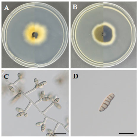
Fig. 1. Morphology of Paradendryphiella arenariae KNUE25A105. A, B: Colonies after 7 days of growth at 25﹎. The left side shows the front view, and the right side shows the reverse view, on malt extract agar (A) and potato dextrose agar (B). C: Conidiophores and conidiogenous cells (Bar=20 µm), D: Conidia (Bar=20 µm).
Diaporthe novem J.M. Santos, Vrandečić & A.J.L. Phillips, Persoonia 27: 14 (2011) [MB#518521] Fig. 2
Morphological characteristics of strain KNUE25A206: On MEA, after 7 days of growth at 25°C in the dark, the colony grew rapidly, reaching 77.0–85.0 mm in diameter. The colony had an irregular shape with a layered, cottony surface that was white and became lighter toward the edges. The reverse side of the colony was white in the center and faded toward the margin. On PDA, the colony reached 72.0–81.0 mm in diameter after 7 days. The colony surface was circular and white, with a dense layered texture and abundant aerial mycelium. The reverse side was light yellow at the center and became lighter toward the outer margin. Under light microscopy, vegetative mycelia were septate and hyaline. Conidia (alpha-conidia) were hyaline, ovoid to ellipsoid, contained two oil droplets (biguttulate), and measured (4.65–)5.35(–6.23) × (2.09–)2.60(–3.08) µm (n = 20).
Specimen examined: Pohang-si, Gyeongsangbuk-do, Korea, 35°59′44.8″N, 129°33′58.1″E, isolated from the internal tissue of Codium fragile, September 2, 2025, strain KNUE25A206 (NIBRFGC000515218), GenBank No. PZ437649 (ITS), and PZ443401 (tef1-α).
Notes: Type strain: CBS 127270. D. novem was first described in 2011 from seeds of Glycine max [21]. Since its original description, this species has been reported in various hosts and geographic regions, including Citrus species in Europe [22,23]. The genus Diaporthe is a species-rich group containing many economically important plant pathogens, endophytes, and saprophytes [22]. The morphological characteristics of the strain KNUE25A206 were similar to those of D. novem [21]. Most of the morphological characteristics of strain KNUE25A206, including aseptate, hyaline, smooth-walled, and ellipsoidal conidia, were consistent with those reported for D. novem. However, the conidia of KNUE25A206 were broadly ellipsoidal and appeared closer to a sub-globose shape than those previously described [21]. However, filiform beta-conidia were not observed in the present study. Colonies grown on PDA exhibited denser and more layered mycelial growth than those grown on MEA.
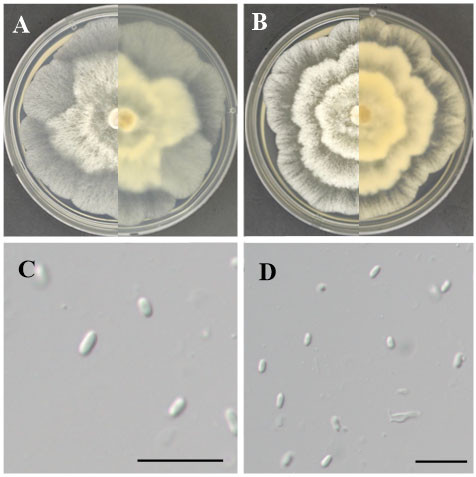
Fig. 2. Morphology of Diaporthe novem KNUE25A206. A, B: Colonies after 7 days of growth at 25﹎. The left side shows the front view, and the right side shows the reverse side, on malt extract agar (A) and potato dextrose agar (B). C & D: Conidia (Bar=20 µm). Only alpha-conidia were observed.
The ITS sequence of strain KNUE25A105 showed 99.64% identity with P. arenariae CBS 181.58 (DQ411539), and the LSU sequence showed 98.89% identity with P. arenariae AFTOL-ID 995 (EU848587). The SSU and rpb2 sequences showed 99.27% and 98.9% identities with CBS 181.58 (KC793336) and AFTOL-ID 995 (DQ470924), respectively. In the ML phylogenetic tree constructed using the combined ITS, LSU, SSU, and rpb2 sequences, strain KNUE25A105 formed a strongly supported monophyletic clade with the ex-type strain P. arenariae CBS 181.58, with 100% bootstrap support (Fig. 3). The ITS sequence of strain KNUE25A206 showed 100% identity with D. novem FP14k-Pod (OP603340), and the tef1-α sequence showed 99.35% identity with D. novem FP14k-Pod (OQ301657). In the ML phylogenetic tree constructed using concatenated ITS and tef1-α sequences, strain KNUE25A206 formed a robust monophyletic group with the reference strain D. novem CBS 127270, with high bootstrap support (Fig. 4).
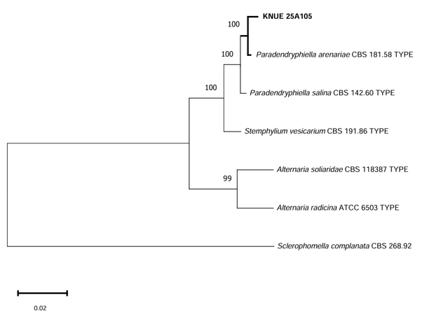
Fig. 3. Maximum likelihood tree of Paradendryphiella arenariae KNUE25A105. The tree is based on concatenated sequences of internal transcribed spacer (ITS), large subunit ribosomal RNA (LSU), small subunit ribosomal RNA (SSU), RNA polymerase II second largest subunit (rpb2) DNA sequences. Sclerophomella complanata was used as an outgroup. The numbers on the nodes represent bootstrap values greater than 50% (1,000 replicates).
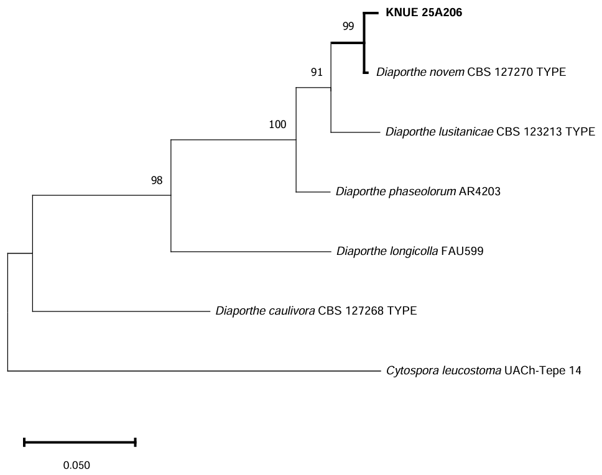
Fig. 4. Maximum likelihood tree of Diaporthe novem KNUE25A206. The tree is based on concatenated sequences of internal transcribed spacer (ITS) and translation elongation factor 1-alpha (tef1-α) DNA sequences. Cytospora leucostoma was used as an outgroup. The numbers on the nodes represent bootstrap values greater than 50% (1,000 replicates).
In this study, Diaporthe novem and Paradendryphiella arenariae were isolated from healthy tissues of the marine green macroalga Codium fragile, collected from the Korean coast [24]. Although P. arenariae has previously been reported in the brown macroalga Agarum clathratum in Korea, its identification was based solely on the ITS region [24]. By employing multi-locus phylogenetic analyses in combination with morphological observations, the present study provides the first detailed morphological and multi-locus phylogenetic characterization of this species from a marine macroalgal host in Korea.
Marine macroalgae harbor diverse fungal communities; however, the fungal diversity associated with green macroalgae remains poorly understood compared to that of terrestrial plants and brown algae [2,3]. Previous studies in Korea have mainly focused on fungal communities associated with brown algae [2]. Therefore, the present study contributes to broadening the known ecological distribution of marine-associated endophytic fungi in Korean coastal environments.
Paradendryphiella arenariae has been previously reported from coastal and marine environments [24]. Recent studies have shown that some isolates of this species may produce biologically active secondary metabolites, including antibacterial compounds and tenuazonic acid [20,25]. Although the ecological role and metabolic characteristics of strain KNUE25A105 remain unclear, these reports suggest that marine-associated isolates of P. arenariae possess diverse metabolic characteristics.
Diaporthe novem is mainly found in terrestrial plant hosts, including soybeans and kiwifruits, where species of Diaporthe are commonly reported as pathogens, endophytes, or saprobes [22]. Several species of the genus Diaporthe have been reported as endophytes in mangrove plants, while others have been isolated from deep-sea sediments [26,27]. The isolation of D. novem from the asymptomatic tissues of C. fragile suggests that marine macroalgae may serve as habitats for phylogenetically diverse fungal taxa. However, the ecological role of this species in marine algal tissues remains unclear and requires further investigation.
Overall, this study provides the first record of P. arenariae and D. novem associated with C. fragile in Korea and provides baseline information for future studies on marine algal-associated fungal diversity.
No potential conflicts of interest relevant to this article were reported.
This work was supported by a grant (NIBR202602103) from the National Institute of Biological Resources (NIBR) funded by the Ministry of Environment (MOE) of the Republic of Korea.
1. Liao C, Doilom M, Jeewon R, Hyde KD, Manawasinghe IS, Chethana KWT, Balasuriya A, Thakshila SAD, Luo M, Mapook A, et al. Challenges and update on fungal endophytes: Classification, definition, diversity, ecology, evolution and functions. Fungal Divers 2025;131:301-67. https://doi.org/10.1007/s13225-025-00550-5
[DOI]
2. Kim JW, Choi YN, Kim EJ, Eom AH. Endophytic fungi isolated from the brown alga Sargassum thunbergii in coastal Korea. Mycobiology 2025;53:794-806. https://doi.org/10.1080/12298093.2025.2578051
[DOI][PubMed][PMC]
3. Sarasan M, Puthumana J, Job N, Han J, Lee JS, Philip R. Marine algicolous endophytic fungi-a promising drug resource of the era. J Microbiol Biotechnol 2017;27:1039-52. https://doi.org/10.4014/jmb.1701.01036
[DOI][PubMed]
4. Chavanich S, Harris LG, Je JG, Kang RS. Distribution pattern of the green alga Codium fragile (Suringar) Hariot, 1889 in its native range, Korea. Aquat Invasions 2006;1:99-108. https://doi.org/10.3391/ai.2006.1.3.1
[DOI]
5. Sarasan M, Job N, Puthumana J, Ravinesh R, Prabhakaran MP, Thomas LC, Philip R. Exploration and profiling of hidden endophytic mycota of marine macroalgae with potential drug leads. FEMS Microbiol Lett 2020;367:fnaa078. https://doi.org/10.1093/femsle/fnaa078
[DOI][PubMed]
6. Erbert C, Lopes AA, Yokoya NS, Furtado NAJC, Conti R, Pupo MT, Lopes JLC, Debonsi HM. Antibacterial compound from the endophytic fungus Phomopsis longicolla isolated from the tropical red seaweed Bostrychia radicans. Bot Mar 2012;55:435-40. https://doi.org/10.1515/bot-2011-0023
[DOI]
7. Kjer J, Debbab A, Aly AH, Proksch P. Methods for isolation of marine-derived endophytic fungi and their bioactive secondary products. Nat Protoc 2010;5:479-90. https://doi.org/10.1038/nprot.2009.233
[DOI][PubMed]
8. Cha JE, Kim YJ, Kim JW, Eom AH. Two new records of endophytic fungi isolated from Lindera obtusiloba in Korea: Colletotrichum citricola and Valsa ceratophora. Kor J Mycol 2024;52:135-43. https://doi.org/10.4489/kjm.520206
[DOI]
9. Eo JK, Choi MS, Eom AH. Diversity of endophytic fungi isolated from Korean ginseng leaves. Mycobiology 2014;42:147-51. https://doi.org/10.5941/MYCO.2014.42.2.147
[DOI][PubMed][PMC]
10. Gardes M, Bruns TD. ITS primers with enhanced specificity for basidiomycetes-application to the identification of mycorrhizae and rusts. Mol Ecol 1993;2:113-8. https://doi.org/10.1111/j.1365-294x.1993.tb00005.x
[DOI][PubMed]
11. White TJ, Bruns TD, Lee SB, Taylor JW. Amplification and direct sequencing of fungal ribosomal RNA genes for phylogenetics. In: Innis MA, Gelfand DH, Sninsky JJ, White TJ, editors. PCR protocols: A guide to methods and applications. San Diego: Academic Press; 1990. p. 315-22. https://doi.org/10.1016/B978-0-12-372180-8.50042-1
[DOI][PubMed][PMC]
12. O’Donnell K. Fusarium and its near relatives. In: Reynolds DR, Taylor JW, editors. The fungal holomorph: Mitotic, meiotic and pleomorphic speciation in fungal systematics. Wallingford: CAB International; 1993. p. 225-33
13. Vilgalys R, Hester M. Rapid genetic identification and mapping of enzymatically amplified ribosomal DNA from several Cryptococcus species. J Bacteriol 1990;172:4238-46. https://doi.org/10.1128/jb.172.8.4238-4246.1990
[DOI][PubMed][PMC]
14. Rehner S. Primers for elongation factor 1-α (EF1-α) [Internet]. Beltsville: Insect Biocontrol Laboratory; 2001 [cited 2025 Apr 30]. Available from: https://web.archive.org/web/20060518132929/http://ocid.NACSE.ORG/research/deephyphae/EF1primer.pdf
15. Liu YJ, Whelen S, Hall BD. Phylogenetic relationships among ascomycetes: Evidence from an RNA polymerase II subunit. Mol Biol Evol 1999;16:1799-808. https://doi.org/10.1093/oxfordjournals.molbev.a026092
[DOI][PubMed]
16. Tamura K, Stecher G, Kumar S. MEGA11: Molecular evolutionary genetics analysis version 11. Mol Biol Evol 2021;38:3022-7. https://doi.org/10.1093/molbev/msab120
[DOI][PubMed][PMC]
17. Nicot J. Une moisissure arénicole du littoral atlantique: Dendryphiella arenaria sp. nov. Rév Mycol 1958;23:87-99.
18. Ellis MB. More dematiaceous hyphomycetes. Kew: Commonwealth Mycological Institute; 1976.
[DOI]
19. Woudenberg JHC, Groenewald JZ, Binder M, Crous PW. Alternaria redefined. Stud Mycol 2013;75:171-212. https://doi.org/10.3114/sim0015
[DOI][PubMed][PMC]
20. Shankar S, Sathiavelu M. Paradendryphiella arenariae an endophytic fungus of Centella asiatica inhibits the bacterial pathogens of fish and shellfish. Front Microbiol 2024;15:1441525. https://doi.org/10.3389/fmicb.2024.1441525
[DOI][PubMed][PMC]
21. Santos JM, Vrandečić K, Cosić J, Duvnjak T, Phillips AJ. Resolving the Diaporthe species occurring on soybean in Croatia. Persoonia 2011;27:9-19. https://doi.org/10.3767/003158511×603719
[DOI][PubMed][PMC]
22. Díaz GA, Latorre BA, Jara S, Ferrada E, Naranjo P, Rodríguez J, Zoffoli JP. First report of Diaporthe novem causing postharvest rot of kiwifruit during controlled atmosphere storage in Chile. Plant Dis 2014;98:1274. https://doi.org/10.1094/PDIS-02-14-0183-PDN
[DOI][PubMed]
23. Guarnaccia V, Crous PW. Emerging citrus diseases in Europe caused by species of Diaporthe. IMA Fungus 2017;8:317-34. https://doi.org/10.5598/imafungus.2017.08.02.07
[DOI][PubMed][PMC]
24. Lee S, Park MS, Lee H, Kim JJ, Eimes JA, Lim YW. Fungal diversity and enzyme activity associated with the macroalgae, Agarum clathratum. Mycobiology 2019;47:50-8. https://doi.org/10.1080/12298093.2019.1580464
[DOI][PubMed][PMC]
25. Kumari A, Singh K, Tiwari N, Katiyar D, Singh SP, Mishra A. Paradendryphiella arenariae (MW504999) as a novel fungal source of tenuazonic acid in tomato (Lycopersicon esculentum). Indian J Microbiol 2025;65:1121-32. https://doi.org/10.1007/s12088-025-01494-9
[DOI][PubMed][PMC]
26. Devadatha B, Jones EBG, Pang KL, Abdel-Wahab MA, Hyde KD, Sakayaroj J, Bahkali AH, Calabon MS, Sarma VV, Sutreong S, Zhang SN. Occurrence and geographical distribution of mangrove fungi. Fungal Divers 2021;106:137-227. https://doi.org/10.1007/s13225-020-00468-0
[DOI]
27. Niu Z, Chen Y, Guo H, Li SN, Li HH, Liu HX, Liu Z, Zhang W. Cytotoxic polyketides from a deep-sea sediment derived fungus Diaporthe phaseolorum FS431. Molecules 2019;24:3062. https://doi.org/10.3390/molecules24173062
[DOI][PubMed][PMC]