Introduction
The freshwater environment consists of various diverse microorganisms and provides plenty of habitat for fungi, such as plant litter, soil, and freshwater plants. In the environment, fungi play a role in controlling nutrient and carbon cycles as a decomposer [1].
The genus Penicillium belongs phylogenetically to Trichocomaceae, and the name is originated from penicillus, little brush by Link. Penicillium is a ubiquitous and widespread fungi, which can live in any environment such as soil, air, indoor and various foods, and produce beneficial secondary metabolite [2]. To our knowledge, over 100 species of Penicillium have been reported in Korea [3]. Recently, P. piscarium from Seungcheon reservoir and P. pasqualens and P. sanguifluum from Dokdo have been reported in Korea [3,4].
In this study, we isolated three Penicillium species from the freshwater environment in Korea. Through molecular phylogenetic analysis and morphological characterization, the three fungal strains were identified as Penicillium guanacastense, P. saturniforme, and P. scabrosum. Here, we present their mycological descriptions and phylogenetic relationships.
Fungal strains were collected from soil underwater at Namsaengi-mot in Jeju, Soyang-river in Chuncheon-si and Gamcheon in Gimcheon-si between 2016-2018 (Table 1). Soil samples were treated using the dilution plate method and then incubated at 20℃. Three-point inoculation was performed on potato dextrose agar (PDA; Difco, BD, Franklin Lakes, NJ, USA), Czapek yeast autolysate agar (CYA; Czapek concentrate 10 mL, sucrose 30 g, yeast extract 5 g, K2HPO4 1 g, trace elements stock solution 1 mL, agar 20 g, dH2O 1 L), yeast extract sucrose agar (YES;yeast extract 20 g, sucrose 150 g, MgSO4∙7H2O 0.5 g, trace element stock solution 1 mL, agar 20 g, dH2O 885 mL) and creatine sucrose agar (CREA; sucrose 30 g, creatine∙1H2O 3 g, K3PO4∙7H2O, MgSO4∙7H2O 0.5 g, KCl 0.5 g, FeSO4∙7H2O 0.01 g, bromocresol purple 0.05 g, trace element stock solution 1 mL, agar 20 g, dH2O 1 L) to observe the morphological characters of the isolated strains [2]. Colony diameter was measured after 7 days of inoculation, and morphological characteristics were observed using a model Eclipse Ni-U microscope (Nikon, Tokyo, Japan).

Fungal genomic DNA was isolated using the NucleoSpin Plant II DNA extraction Kit (Macherey-Nagel, Duren, Germany). For the identification of the fungi, amplification of an internal transcribed spacer (ITS) region using primers ITS1 (5’-TCCGTAGGTGAACCTGCGG-3’) and ITS4 (5’-TCCTCCGCTTATTGATATGC-3’), ß-tubulin gene using Bt2a (5’-GGTAACCAAATCGGTGCTGCTTTCG-3’) and Bt2b (5’-ACCCTCAGTGTAGTGACCCTTGGCG-3’) and calmodulin gene using cmd5 (5’-CCGAGTACAAGGARGCCTTC-3’) and cmd6 (5’-CCGATRGAGGTCATRACGTGG-3’) were performed [5]. Similarity searches of the DNA sequences were carried out using BLASTn algorithms available at the National Center for Biotechnology Information (NCBI). For the phylogenetic analysis, MEGA7 software was used [6]. A phylogenetic tree was constructed by the neighbor-joining (NJ) method with 1,000 bootstrap replications. Reference sequences of other fungi were obtained from GenBank at NCBI (Table 2).
Table 2. GenBank accession numbers of isolates includes in this study
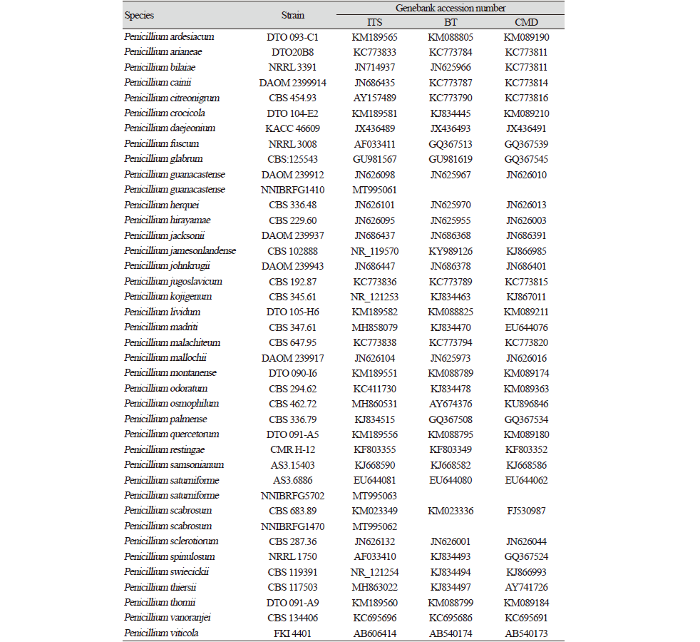
|
|
ITS: Internal transcribed spacer; BT: ß-tubulin gene; CMD: Calmodulin gene |
Taxonomy
Penicillium guanacastense K.G. Rivera, Urb & Seifert, Mycotaxon 119, 315-28 (2012) [7] (Fig. 1 and 2).
Mycobank No.: MB563044
Description:
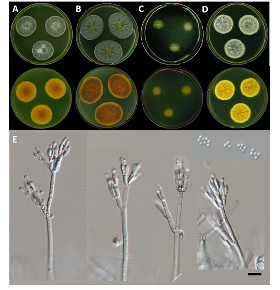
Colonies on PDA texture floccose to granular, slight radial sulcate, front olive green to dark green, reverse dark brown to orange, 4.5 mm on 7 days at 25℃; YES texture velutinous to fasciculate, sulcate, some yellow mycelium, front dark green to pale grayish green, reverse light green to light yellow, 5 mm on 7 days at 25℃; CYA texture velutinous, exudate present orange droplet, radial sulcate, front center white, middle whitish green, margin light lemon, reverse dark to light orange, 4 mm on 7 days at 25℃; CREA high acid production.
The conidiophore monoverticillate, smooth-walled, short stipes, hyaline; phialide 9.3-12 μm length×3.8-4.46 μm width, cylindrical; conidia 2.77-4.47 μm diameter, globose to subglobose.
Phylogenetic section: Sclerotiora

Fig. 1.Phylogenetic analysis using the neighbor-joining method based on internal transcribed spacer (ITS) region, ß-tubulin and calmodulin gene sequences of three Penicillium species. P. consobrinum was used to an outgroup. Bootstrap values more than 50% (1,000 replications) are shown at branches. The new isolates from the present study are shown in bold and red.
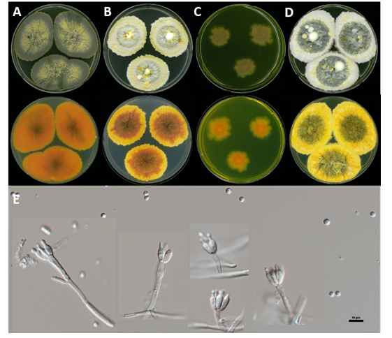
Habitat: Soil in stream
Specimen examined: Namsaengi-mot, Chocheon-uep, Jeju-do, Republic of Korea, 22 Mar 2016, NNIBRFG1410, Nakdonggang National Institute of Biological Resources
Note: NNIBRFG1410 is phylogenetically closely related to P. guanacastense within Sclerotiora clade. The molecular data of NNIBRFG1410 is identical to the strain of P. guanacastense,with high similarity in the ITS, ß-tubulin and calmodulin sequence data, respectively. We confirm NNIBRFG1410 as P. guanacastense based on the morphology and phylogeny, and report the freshwater environment. The morphology of NNIBRFG1410 and Ref. species are similar conidiophore, philade and conidia (Table 3).
Table 3. The morphology of Penicillium species.
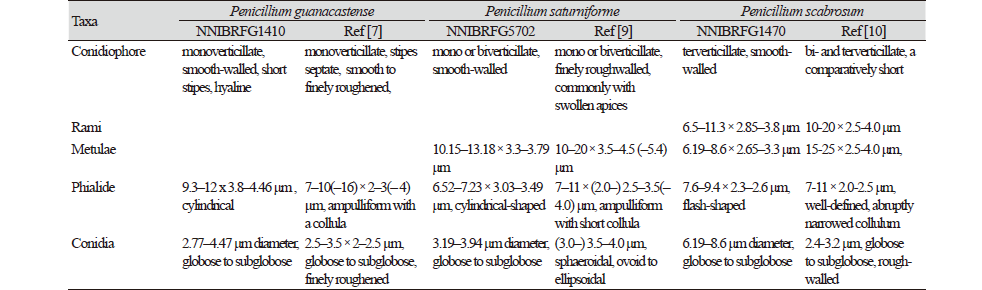
|
|
ITS: Internal transcribed spacer; BT: ß-tubulin gene; CMD: Calmodulin gene |
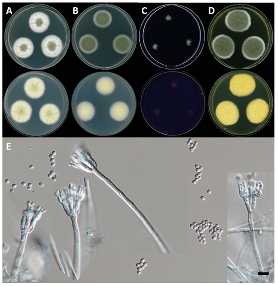
Penicillium saturniforme (L. Wang & W.Y. Zhuang) Houbraken & Samson, Studies in Mycology 70: 48 (2011)[8] (Fig. 1and 3).
≡EuPenicillium saturniforme L. Wang & W.Y. Zhuang, Mycopathologia 167 (6): 300 (2009).
Mycobank No.: MB541663
Description:
Colonies on PDA texture velutinous, radial slightly sulcate, front olive green to white, reverse pale yellow, 3 mm on 7 days at 25℃; YES texture floccose to fasciculate, slightly radial sulcate, front dark green to white, reverse light yellow, 3.5 mm on 7 days at 25℃; CYA texture floccose to granular, front dark green to white, reverse pale yellow, 2.5 mm on 7 days at 25℃
The conidiophore mono or biverticillate, smooth-walled; metulae 10.15-13.18 μm length×3.3-3.79 μm width; phialide 6.52-7.23 μm length×3.03-3.49 μm width, cylindrical-shaped; conidia 3.19-3.94 μm diameter, globose to subglobose.
Phylogenetic section: Aspergilloides
Habitat: Soil in the river
Specimen examined: Soyang-river, Chuncheon-si, Gangwon-do, Republic of Korea, 03 May 2018, NNIBRFG5702, Nakdonggang National Institute of Biological Resources
Note: NNIBRFG5702 is phylogenetically closely related to P. saturniforme within Aspergilloides clade. The molecular data of NNIBRFG5702 is identical to the strain of P. saturniforme,with high similarity in the ITS, ß-tubulin and calmodulin sequence data, respectively. We confirm NNIBRFG5702 as P. saturniforme based on the morphology and phylogeny, and report the freshwater environment. The morphology of NNIBRFG5702 and Ref. species are similar conidiophore, matulae, philade and conidia (Table 3).
Penicillium scabrosum Frisvad, Samson & Stolk, Persoonia 14 (2): 177 (1990)[9] (Fig. 1 and 4).
Mycobank No.: MB136735
Description:
Colonies on PDA texture floccose to fasciculate, radial sulcate, front pale green to dark green, reverse dark orange to light orange, 2.5 mm on 7 days at 25℃; YES texture floccose to granular, deeply radial sulcate, front grayish green to light moss green, reverse yellow, 2.5 mm on 7 days at 25℃; CYA texture floccose to granular, deeply radial sulcate, exudate pale yellow droplet, front light green, reverse dark grayish orange, 3.5 mm on 7 days at 25℃; CREA good acid production
The conidiophore terverticillate, smooth-walled; Rami 6.5-11.3 μm length×2.85-3.8 μm width; metulae 6.19-8.6 μm length×2.65-3.3 μm width; phialide, 7.6-9.4 μm length×2.3-2.6 μm width, flash-shaped; conidia 6.19-8.6 μm diameter, globose to subglobose.
Phylogenetic section: Ramosa
Habitat: Soil in the stream
Specimen examined: Gamcheon, Gimcheon-si, Gyeongsangbuk-do, Republic of Korea, 23 Mar 2016, NNIBRFG1470, Nakdonggang National Institute of Biological Resources
Note: NNIBRFG1470 is phylogenetically closely related to P. scabrosum within Ramosa clade. The molecular data of NNIBRFG1470 is identical to the strain of P. scabrosum,with high similarity in the ITS, ß-tubulin and calmodulin sequence data, respectively. We confirm NNIBRFG1470 as P. scabrosum based on the morphology and phylogeny, and report the freshwater environment. The morphology of NNIBRFG1470 and Ref. species are similar conidiophore, rami, matulae and philade. Conidia size of NNIBRFG1470 are bigger than Ref. species (Table 3).

