INTRODUCTION
Freshwater fungi are taxonomically diverse and a polyphylogenetic group that is found in aquatic or semi-aquatic environments [1]. Well-known habitats of freshwater fungi include soil, plant litter such as submerged wood and leaves, and plants and animals of which the exist as endophytes [2]. The main ecological role of freshwater fungi is the degradation of organic materials in water, including plant litter and/or dead animal tissues. Some of these fungi are pathogens that can cause plant diseases or endophytes living symbiotically on plant tissues [3]. However, many of the biological roles of fungi in aquatic ecosystems, and their physiological and biochemical characteristics remain undetermined. Four fungal genera relative to the current study are Nothophoma, Westerdykella, Chrysosporium, and Taeniolella.
The genus Nothophoma belongs to Didymellaceae, one of the largest families within the order Pleosporales of the phylum Ascomycota. To date, 21 species have been classified under genus [4]. Many species of this genus are reported to be plant pathogens [5]. Members of this genus are characterized by anamorphic stage and conidia morphology [6].
The genus Westerdykella belongs to Sporormiaceae, one of the families in the order Pleosporales. To date, 18 species belonging to this genus have been identified with many being reported to be saprobic isolates from dung [7,8]. Westerdykella species are known to be a potential bioresource capable of producing various bioactive substances [7]. Westerdykella species are characterized by their black globose astomatous ascomata, multi-spored asci, and brown ascospores [9].
The genus Chrysosporium belongs to Onygenaceae, one of the largest families of the Ascomycota order Onygenales. Currently, more than 60 species belonging to this genus have been reported [10]. Some species of this genus are isolated from human and animal skin, and exhibited keratinophilic activity [11]. In addition, this genus is commonly found in soils. This genus is classified according to characteristics of an anamorphic stage and its conidia morphology [12].
The genus Taeniolella belongs to Mytilinidiaceae, which is the largest family of Ascomycota order Mytilinidiales. More than 20 species belonging to this genus have been reported to date [13]. Fungal species belonging to this genus come from saprobes lichenicolous taxa and demonstrate a wide range of habits and ecological preferences [14]. Taeniolella is classified based on its morphology of asexual stage - conidiophores, holoblastic conidiogenous cells, and aseptate to pluriseptate conidia formed in acropetal chains [15].
For the first time, we isolated seven subphylum Pezizomycotina species from environmental samples, Nothophoma spiraeae, Westerdykella aquatica, W. aurantiaca, W. dispersa, Chrysosporium sanyaense, C. pseudomerdarium and Taeniolella phialosperma. The environmental samples included submerged plant litter, and sediment from freshwater environments including rivers, wetland, and streams. Molecular phylogenetic and morphological characteristics of the isolates were investigated.
MATERIALS AND METHODS
Isolation of fungal strains and culture conditions
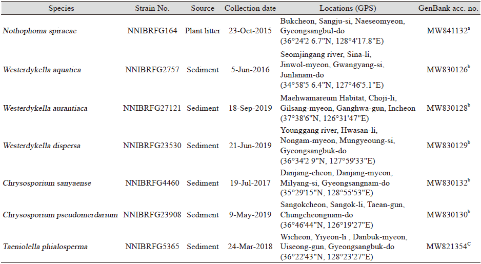
Fungal strains were collected from plant litter and sediment sampled from freshwater environment. The collection information of all strains identified in this study is listed in Table 1. To isolate fungal strains, plant litter samples were washed with distilled water at least twice, and incubated in a pretreatment liquid medium (0.05% 3-morpholinopropane-1-sulfonic acid [weight/volume (w/v)], 0.05% KNO3 [w/v], 0.025% KH2PO4 [w/v], and 0.025% K2HPO4 [w/v]) at 20℃ for three days. Then, 100 µL of the pretreatment medium was spread on a 1% water agar plate and incubated at 20℃ for two days. Hyphal tips and germinated conidia were isolated under a microscope and transferred onto a 24-well plate containing V8 agar (V8A; 8% V8 juice [v/v] and 1.5% agar [w/v], adjusted to pH 6.0 using 10 N NaOH) and incubated at 25℃ in the dark. To isolate fungal strains from sediments, a dilution method was used. Diluted suspension (200 μL) of freshwater sediment and distilled water (1:200 and 1:2,000) were spread on potato dextrose agar (PDA; 3.9% potato dextrose agar powder [w/v]; Difco, Sparks, MD, USA) containing 50 ppm streptomycin, and fungal strains were isolated in the pure form after incubation for 4-5 days at 25℃ by repeating this step. All strains identified in this study were grown on malt extract agar (MEA; 2% malt extract [w/v] and 2% agar [w/v]), oatmeal agar (OA; 7.25% oatmeal agar powder [w/v]; Difco, Sparks, MD, USA), potato carrot agar (PCA; 2.4% potato carrot agar powder [w/v]; HiMedia, Mumbai, India), corn meal agar (CMA; 3.9% corn meal agar powder [w/v]; Difco, Sparks, MD, USA), Czapek-dox solution agar (CDA; Difco Sparks, MD, USA), dichloran glycerol chloramphenicol agor (DG18A; Merck Millipore, Billerica, MA, USA), and corn meal dextrose agar (CMDA; 2% cornmeal [w/v], 2% glucose [w/v], and 2% agar), and yeast extract peptone dextrose agar (YPDA; Duchefa Biochemie, Haarlem, the Netherlands). All strains were preserved at Nakdonggang National Institute of Biological Resources (NNIBR), Sangju, Korea
Table 1. Information of strains used in this study.
|
|
aBeta tubulin (TUB), bInternal transcripbed spacer (ITS), cLarge subunit of ribosomal DNA (LSU). |
Morphological analysis
Microstructures of fungal species were observed under an Eclipse Ni light microscope (Nikon, Tokyo, Japan) equipped with a Ds-Ri2 digital camera (Nikon, Tokyo, Japan). At least 50 individuals were examined for observation and measurement of each structure.
DNA extraction, polymerase chain reaction (PCR), and DNA sequencing
Fungal genomic DNA was isolated using a NucleoSpin® Plant II DNA extraction kit (Macherey-Nagal, Düren, Germany). For molecular identification of the fungi, PCR amplifications were performed for the internal transcribed spacer (ITS) rDNA regions using primers ITS1 (5′-TCCGTAGGTGAACCTGCGG-3′) and ITS4 (5′-TCCTCCGCTTATTGATATGC-3′) [16], for the large subunit of rDNA (LSU) using primers LROR (5′-ACCCGCTGAACTTAAGC-3’) and LR7 (5′-TACTACCACCAAGATCT-3′) [17], for the beta tubulin gene (TUB) using primers bt2a (5′-GGTAACCAAATCGGTGCTGCTTTC-3′) and bt2b (5′-ACCCTCAGTGTAGTGACCCTTGGC-3′) [18]. Amplicons were sequenced by a DNA sequencing service (Macrogen Inc., Daejeon, Korea) using the same primers as those used for amplification. A homology search of the DNA sequences was performed using the BLAST algorithms available from the National Center for Biotechnology Information (NCBI; https://www.ncbi.nlm.nih.gov).
Phylogenetic analysis
We obtained the sequences of the reference species from NCBI for phylogenetic analyses, which are shown in Fig. 1-4. All reference sequences have been reported previously [5-8,10,11,13,14,19,20]. Sequences were edited using the DNAStar software package version 5.05 (DNAStar, Inc., Madison, WI, USA). The accession numbers of the sequences used in this study are shown in the phylogenetic trees (Fig. 1-4 and Table 1) constructed using the maximum likelihood (ML) method. ML analysis was performed using MEGA 7.1 [21] with the default settings of the program, except for replacement with the Tamura-Nei model. Bootstrapping analysis of 1,000 replicates was performed to test the robustness of each grouping.
RESULTS AND DISCUSSION
Phylogenetic analysis
Phylogenetic analysis was performed to identify the seven fungal strains and infer their phylogenetic relationships to other similar species. TUB sequences were used for phylogenetic analysis of Nothophoma and its related species. As Fig. 1, strain NNIBRFG164 formed a clade with three strains of N. spiraeae including the type material. BLASTn analysis of the TUB gene of NNIBRFG164 showed a high similarity of 99.69% with that of N. spiraeae. ITS sequences were used for the phylogenetic analysis of Westerdykella (Fig. 2). Strain NNIBRFG2757 and NNIBRFG2985 formed a clade with two W. aquatica strains, including the type material. NNIBRFG27121 formed a clade with two W. aurantiaca strains. Another strain, NNIBRFG23530, formed a clade with two W. dispersa strains, including the type material. BLASTn analysis of the ITS of NNIBRFG2757 revealed a high similarity of 99.78% with that of W. aquatica strain JAUCC1788. The ITS of NNIBRFG27121 showed a 99.58% similarity with that of W. aurantiaca strain IMI 086825. The ITS of NNIBRFG23530 showed a 99.82% similarity with that of W. dispersa strain CBS 297.56.
ITS sequences were used for phylogenetic analysis Chrysosporium and its related species. As shown in Fig. 3, strains NNIBRFG2776 and NNIBRFG4460 formed a clade with C. sanyaense and NNIBRFG23908 formed a clade with C. pseudomerdarium. BLASTn analysis showed the ITS of NNIBRFG2776 and NNIBRFG4460 had a 99.82% similarity with that of C. sanyaense strain GZUIFR-A10222M while the ITS of NNIBRFG23908 had a 100% similarity with that of C. pseudomerdarium strain IJL01.
The LSU sequences were used for phylogenetic analysis of Taeniolella and its related species. Strain NNIBRFG5365 formed a clade with three isolates of T. phialosperma including, the type material (Fig. 4). The LSU of NNIBRFG5365 exhibited 100% similarity with that of T. phialosperma.
Taxonomy
Nothophoma spiraeae L.X. Zhang & X.L. Fan, 2020 [MB#833556] (Fig. 1 and Fig. 5)
Description: Colonies grew slightly fast and reached 57 mm on PDA, 67 mm on PCA, 38 mm on YPDA, 54 mm on OA, 46 mm on MEA, 28 mm on DG18A, 45 mm on CDA, and 46 mm on CMA at 25℃, 10 days post inoculation. The colony color was white with floccose aerial mycelium, turning to olive-green from the center on PDA; hyaline white with short aerial mycelium turning to dark-green near the center on MEA; creamy white with fluffy aerial mycelium on OA; creamy white with cottony aerial mycelium on PCA; dark-green with a smooth surface on CMA; light gray with cottony aerial mycelium on CDA; light yellow with dense aerial mycelium on DG18A; and goldenrod with a dense smooth surface on YPDA. The optimal growth conditions were 25℃ on OA media. Conidia were ovoid, oblong to ellipsoidal, smooth, thin-walled, aseptate, rounded at both ends, hyaline to olivaceous buff, and measured 3.6-6.7 µm×2.1-4.1 µm (x=4.82±0.61 µm×3.07±0.42 µm, length (L)/width (W) ratio=1.56, n=50).
Habitat: Plant litter in stream
Specimen examined: Bukcheon, Sangju-si, Naeseomyeon, Gyeongsangbuk-do, Republic of Korea; October 23, 2015; NNIBRFG164
Notes: Nothophoma spiraeae was first reported as a plant pathogen causing canker disease in Spiraea salicifolia [19]. In this study, NNIBRFG164 was isolated from plant litter in stream.
Westerdykella aquatica H.Y. Song & D.M. Hu, Mycotaxon 135 (2): 287 (2020) [MB#825645] (Fig. 2 and Fig. 6)
Description: Colonies grew slightly slow and reached 35 mm on PDA, 32 mm on CMDA, 31 mm on YPDA, 39 mm on V8A, 37 mm on OA, and 38 mm on MEA at 25℃, 7 days post inoculation. The colony color was hyaline with a smooth mycelium surface on CMDA; hyaline with unclear aerial mycelium margins on MEA; creamy white to lemon from the center with fluffy aerial mycelium on OA; creamy white to lemon from center with cottony aerial mycelium on PDA; creamy white to lemon from the center with cottony aerial mycelium on V8A; and white with fluffy aerial mycelium on YPDA. Cleistothecium was observed on PDA at 25℃, 2 weeks post inoculation. Asci were globose to subglobose, and measured 12.2-24.4 µm×11.1-19.7 µm (x=18.2±2.59 µm×14.8±2.21 µm, L/W ratio=1.22, n=50). Ascospore segments were ellipsoidal, subhyaline to light brown, aseptate, measuring 4.6-7.1 µm×2.0-3.6 µm (x=6.08±0.53 µm×2.78±0.39 µm, L/W ratio=2.19, n=50).
Habitat: Sediment in freshwater
Specimens examined: Seomjingang River, Sina-li, Jinwol-myeon, Gwangyang-si, Junlanam-do, Republic of Korea; June 5, 2016; NNIBRFG2757 / Sujangdong-wetland, Panpo-li, Hangyeong-myeon, Jeju-si, Jeju-do, Republic of Korea; August 10, 2016; NNIBRFG2985
Note: The type material of W. aquatica, JAUCC1788, was isolated from mud in a rice field [7]. In this study, NNIBRFG2757 was isolated from sediment in river. Other specimens identified as W. aquatica have been isolated in many local places. For example, NNIBRFG2985 was isolated from aquatic plant root in Jeju Island and NNIBRFG22735 from sediment of Sangju Gonggeumji. Most W. aquatica strains in NNIBR were isolated from sediment of freshwater -streams, lakes, reservoirs, etc.
Westerdykella aurantiaca (J.N. Rai & J.P. Tewari) Kruys, Systematics and Biodiversity 7: 476 (2009) [MB#543081] (Fig. 2 and Fig. 7)
Description: Colonies grew slightly fast and reached 51 mm on PDA, 54 mm on CMDA, 47 mm on YPDA, 48 mm on V8A, 42 mm on OA, and 55 mm on MEA at 25℃, 7 days post inoculation. The colony color was hyaline with a smooth aerial mycelium surface on CMDA; hyaline with smooth aerial mycelium surface on MEA; creamy white to light goldenrod from center with fluffy aerial mycelium on OA; creamy white to lemon from center with fluffy aerial mycelium on PDA; creamy white from center with cottony aerial mycelium on V8A; and creamy white to light yellow with fluffy aerial mycelium on YPDA. Cleistothecium were observed on PDA at 25℃, 2 weeks post inoculation. Asci were globose to subglobose, and measured 12.2-24.4 µm×11.1-19.7 µm (x=18.2±2.59 µm×14.8±2.21 µm, L/W ratio=1.22, n=50). Ascospore segments were ellipsoidal, subhyaline to light brown, aseptate, and measured 4.6-7.1 µm×2.0-3.6 μm (x=6.08±0.53 µm×2.78±0.39 μm, L/W ratio=2.19, n=50).
Habitat: Sediment in wetland
Specimen examined: Maehwamareum Habitat, Choji-li, Gilsang-myeon, Ganghwa-gun, Incheon, Republic of Korea; September 18, 2019; NNIBRFG27121
Note: Westerdykella aurantiaca was first reported as an isolate from soil in India [22]. In this study, NNIBRFG27121 was isolated from wetland sediment.
Westerdykella dispersa (Clum) Cejp & Milko, Ceská Mykologie 18 (2): 83 (1964) [MB#341019] (Fig. 2 and Fig. 8)
Description: Colonies grew slightly fast and reached 41 mm on PDA, 46 mm on CMDA, 48 mm on YPDA, 35 mm on V8A, 37 mm on OA, and 50 mm on MEA at 25℃, 7 days after inoculation. The colony color was hyaline with a smooth aerial mycelium surface on CMDA; hyaline with smooth aerial mycelium surface on MEA; creamy white to light yellow from the center with fluffy aerial mycelium on OA; creamy white to orange from the center with fluffy aerial mycelium on PDA; creamy white to goldenrod from the center with cottony aerial mycelium on V8A; and creamy white to light yellow with fluffy aerial mycelium on YPDA. Cleistothecium was observed on PDA at 25℃, 2 weeks post inoculation. Asci were globose to subglobose, and measured 11.0-13.6 µm×8.6-12.8 µm (x=12.3±0.73 µm×10.8±0.98 µm, L/W ratio=1.13, n=25). Ascospore segments were ellipsoidal, subhyaline to light brown, aseptate, and measured 3.1-4.9 µm×1.6-3.3 µm (x=4.19±0.42 µm×2.42±0.33 µm, L/W ratio=1.17, n=50).
Habitat: Sediment in freshwater
Specimen examined: Younggang river, Hwasan-li, Nongam-myeon, Mungyeoung-si, Gyeongsangbuk-do, Republic of Korea; October 13, 2019; NNIBRFG23530
Note: Westerdykella dispersa was first reported as a mangrove mud isolate from in India [23]. In this study, NNIBRFG233530 was isolated from sediment in river.
Chrysosporium pseudomerdarium Oorschot, Studies in Mycology 20: 14 (1980) [MB#118498] (Fig. 3 and Fig. 9)
Description: Colonies grew slowly and reached 7 mm on PDA, 10 mm on CMDA, 9 mm on YPDA, 11 mm on V8A, 15 mm on OA, and 11 mm on MEA at 25℃, 7 days post inoculation. The color of the colony was hyaline with a short aerial mycelium surface on CMDA; hyaline with short aerial mycelium surface on MEA; ivory to light brown from the center with dense aerial mycelium on OA; creamy white to light brown from the center with short aerial mycelium on PDA; light gray to goldenrod from the center with cottony aerial mycelium on V8A; and creamy white to light yellow with dense aerial mycelium on YPDA. Terminal and lateral conidia were subhyaline, subglobose, pyriform or obovoid, aseptate, thick-walled, and measured 4.7-7.5 µm×3.5-5.9 µm (x=5.9±0.68 µm×4.5±0.49 µm, L/W ratio=1.31, n=50). Intercalary conidia were solitary or catenate, subhyaline, subglobose, pyriform or obovoid, and measured 3.8-6.7 µm×2.8-5.0 µm (x=5.1±0.66 µm×3.8±0.50 µm, L/W ratio=1.32, n=50).
Habitat: Sediment in freshwater
Specimen examined: Sangokcheon, Sangok-li, Taean-gun, Chungcheongnam-do, Republic of Korea; May 9, 2019; NNIBRFG23908
Note: The type material of C. pseudomerdarium was isolated from the lungs of rodents [24]. In this study, NNIBRFG23908 was isolated from sediment in stream.
Chrysosporium sanyaense Yan W. Zhang, Y.F. Han, J.D. Liang & Z.Q. Liang, Mycosystema 32 (4): 613 (2013) [MB#800004] (Fig. 3 and Fig. 10)
Description: Colonies grew slightly fast and reached 34 mm on PDA, 45 mm on CMDA, 33 mm on YPDA, 46 mm on V8A, 45 mm on OA, and 40 mm on MEA at 25℃, 7 days post inoculation. The color of the colony was hyaline with a short aerial mycelium surface on CMDA; hyaline with a short aerial mycelium surface on MEA; ivory to light brown from the center with dense aerial mycelium on OA; creamy white to light brown from the center with short aerial mycelium on PDA; light gray to goldenrod from the center with cottony aerial mycelium on V8A; and creamy white with dense aerial mycelium on YPDA. Terminal and lateral conidia were subhyaline to brown, subglobose, and measured 5.0-8.9 µm×3.0-5.2 µm (x=7.0±0.84 µm×4.3±0.44 µm, L/W ratio=1.64, n=50). Intercalary conidia were subhyaline, subglobose, and measured 3.5-7.6 µm×2.3-5.9 µm (x=5.5±1.10 µm×4.1±0.91 µm, L/W ratio=1.33, n=50).
Habitat: Sediment in stream
Specimens examined: Yujeon-cheon, Daebyeong-myeon, Hapcheon-gun, Gyeongsangnam-do, Republic of Korea; June 6, 2016; NNIBRFG2776/ Danjang-cheon, Danjang-myeon, Milyang-si, Gyeongsangnam-do, Republic of Korea; July 19, 2017, NNIBRFG4460
Note: The type material of C. sanyaense was isolated from palm rhizosphere soil in China [20]. In this study, two strains were isolated from sediment in stream.
Taeniolella phialosperma Ts. Watan., Mycologia 84: 478 (1992) [MB#358145] (Fig. 4 and Fig. 11)
Description: Colonies grew very fast and reached 75 mm on PDA, 69 mm on CMDA, 75 mm on YPDA, 62 mm on V8A, 51 mm on OA, and 69 mm on MEA at 25℃, 7 days post inoculation. The colony color was olive to dark olive green with fluffy aerial mycelium on CMDA; lemon to gray with dense aerial mycelium on MEA; ivory to goldenrod from the center with dense aerial mycelium on OA; dark yellow to dark green from the center with dense aerial mycelium on PDA; olive drab to dark slate gray from the center with dense aerial mycelium on V8A; and ivory with wrinkled aerial mycelium on YPDA. Taeniolella state was observed after three days of culture on media. Phragmospores were holoblastic, brown to dark brown, clavate, ellipsoidal, or cylindrical, multiseptated, and measured 63.2-305.9 µm×12.6-20.6 µm (x=145.0±46.01 µm×17.0±2.15 µm, L/W ratio=8.55, number of septa=8.95, n=50).
Habitat: Sediment in freshwater
Specimen examined: Wicheon, Yiyeon-li, Danbuk-myeon, Uiseong-gun, Gyeongsangbuk-do, Republic of Korea; October 10, 2018; NNIBRFG5365
Note: Taeniolella phialosperma was first reported as an isolate from strawberry rhizosphere soil and paddy field soil in Japan [15]. In this study, NNIBRFG27121 was isolated from sediment in stream.
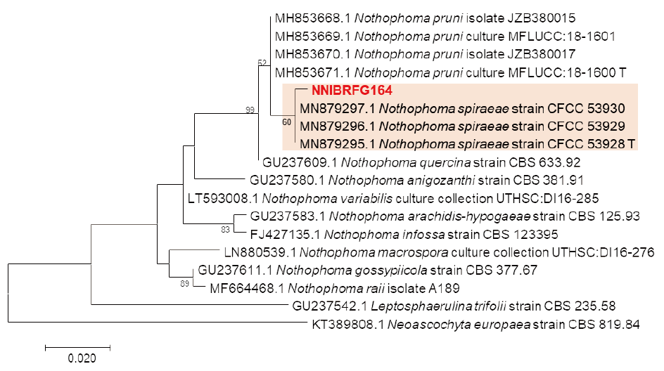
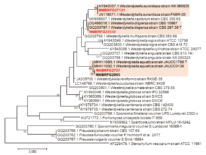
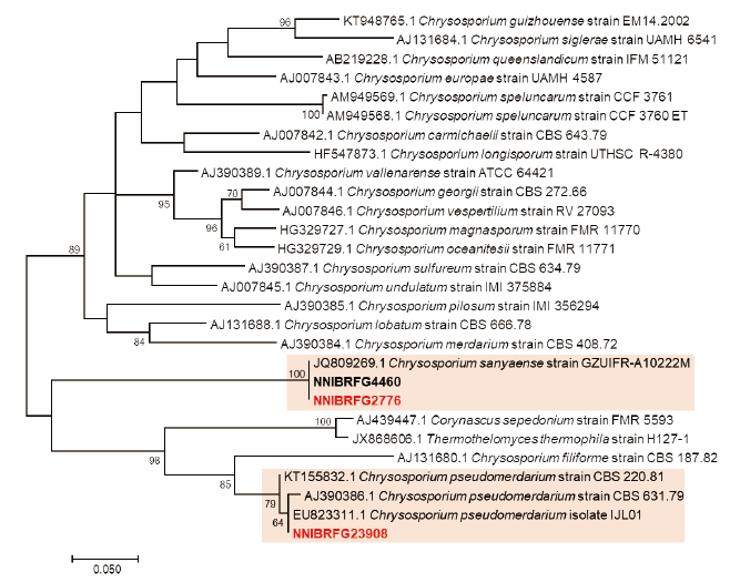
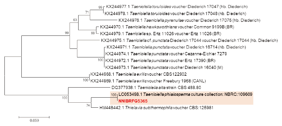

Fig. 5. Morphological characters of Nothophoma spiraeae NNIBRFG164. Mycelial growth on PDA (A), MEA (B), OA (C), PCA (D), CMA (E), CDA (F), DG18 (G) and YPDA (H) for 10 days at 25℃. Graph of growth rate at different temperature were (I). Microscopic observation showed conidia (J) (Scale bars: 20 μm). PDA, potato dextrose agar; MEA, malt extract agar; OA, oatmeal agar; PCA, potato carrot agar; CMA, corn meal dextrose agar; CDA, czapek-dox solution agar; DG18A, dichloran glycerol chloramphenicol; YPDA, yeast extract peptone dextrose agar.

Fig. 6. Morphological characters of Westerdykella aquatica NNIBRFG2757. Mycelial growth on CMDA (A), PDA (B), MEA (C), V8A (D), OA (E), YPDA (F) for 7 days at 25℃. Microscopic observation showed ascospore segment (G) and asci (H) (Scale bars: 10 μm). CMDA, corn meal dextrose agar; PDA, potato dextrose agar; MEA, malt extract agar; V8A, V8 juice agar; OA, oatmeal agar; YPDA, yeast extract peptone dextrose agar.
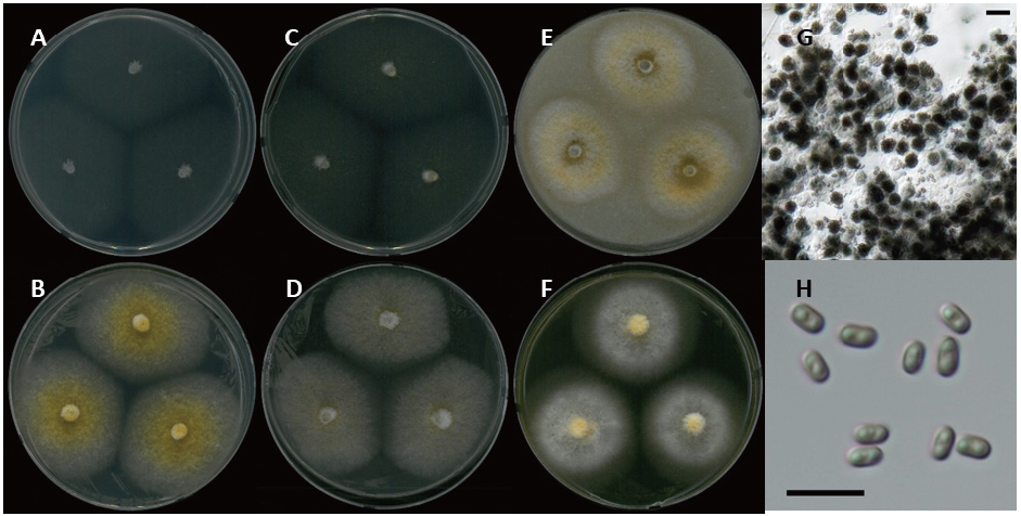
Fig. 7. Morphological characters of Westerdykella aurantiaca NNIBRFG27121. Mycelial growth on CMDA (A), PDA (B), MEA (C), V8A (D), OA (E), YPDA (F) for 7 days at 25℃. Microscopic observation showed asci (G) and ascospore segment (H) (Scale bars: 10 μm). CMDA, corn meal dextrose agar; PDA, potato dextrose agar; MEA, malt extract agar; V8A, V8 juice agar; OA, oatmeal agar; YPDA, yeast extract peptone dextrose agar.

Fig. 8. Morphological characters of Westerdykella dispersa NNIBRFG23530. Mycelial growth on CMDA (A), PDA (B), MEA (C), V8A (D), OA (E), YPDA (F) for 7 days at 25℃. Microscopic observation showed asci (G) and ascospore segment (H) (Scale bars: 10 μm). CMDA, corn meal dextrose agar; PDA, potato dextrose agar; MEA, malt extract agar; V8A, V8 juice agar; OA, oatmeal agar; YPDA, yeast extract peptone dextrose agar.

Fig. 9. Morphological characters of Chrysosporium pseudomerdarium NNIBRFG23908. Mycelial growth on CMDA (A), PDA (B), MEA (C), V8A (D), OA (E), YPDA (F) for 7 days at 25℃. Microscopic observation showed conidia morphology (G, H) (Scale bars: 10 μm). CMDA, corn meal dextrose agar; PDA, potato dextrose agar; MEA, malt extract agar; V8A, V8 juice agar; OA, oatmeal agar; YPDA, yeast extract peptone dextrose agar.
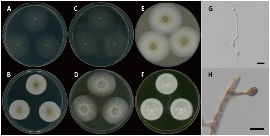
Fig. 10. Morphological characters of Chrysosporium sanyaense NNIBRFG4460. Mycelial growth on CMDA (A), PDA (B), MEA (C), V8A (D), OA (E), YPDA (F) for 7 days at 25℃. Microscopic observation showed conidia morphology (G, H) (Scale bars: 10 μm). CMDA, corn meal dextrose agar; PDA, potato dextrose agar; MEA, malt extract agar; V8A, V8 juice agar; OA, oatmeal agar; YPDA, yeast extract peptone dextrose agar.

Fig. 11. Morphological characters of Taeniolella phialosperma NNIBRFG5365. Mycelial growth on CMDA (A), PDA (B), MEA (C), V8A (D), OA (E), YPDA (F) for 7 days at 25℃. Microscopic observation showed conidia morphology (G, H) (Scale bars: 10 μm). CMDA, corn meal dextrose agar; PDA, potato dextrose agar; MEA, malt extract agar; V8A, V8 juice agar; OA, oatmeal agar; YPDA, yeast extract peptone dextrose agar.

