때죽나무(Styrax japonicus Siebold & Zucc.)는 우리나라와 중국, 일본, 대만에 천연 분포하고, 우리나라에는 중남부지방에 널리 분포한다[1,2]. 꽃, 수피, 수형이 아름답고 각종 공해와 병충해, 해풍과 추위에도 강한 수종으로 평가받아 외국에서 30여 품종이 개발되었으며 가로수, 공원수, 조경수로 널리 식재되고 있다[3]. 우리나라의 때죽나무는 특히 내한성과 내조성(耐潮性, salt spray tolerance)이 강한 편이고, 꽃의 향기가 좋고 꿀이 많아서 밀원수종으로도 이용된다[4,5]. 지금까지 국내의 때죽나무에서 병원균으로 기록된 곰팡이는 흰가루병균(Oidium sp.)과 점무늬병균(Cercoramularia koreana, Pseudocercospora fukuokaensis), 녹병균(Pucciniastrum styracinum) 등이 있다[6,7].
그 중 때죽나무 녹병균은 임업시험장(현, 국립산림과학원)의 1967년 시험연구보고서(상편)에 학명만 기록되어 있을 뿐이며, 상세한 균학적 특징이나 동정의 근거가 되는 측정치 및 특성에 관해서는 전혀 기록되지 않았다[8]. 한편, 녹병에 감염된 때죽나무는 심한 조기낙엽으로 생육이 크게 위축되며 관상가치를 잃는다. 따라서, 관상 조경수로서 뿐만 아니라 공해에 강하여 도시지역의 가로수 및 환경수로서 가치가 높은 수종인 때죽나무의 녹병균에 관하여 상세하게 기록하고, ITS 및 LUS 영역의 염기서열을 제공함으로써 녹병의 진단 및 녹병균의 동정에 기여하고자 한다.
고려대학교 진균표본보관소(KUS)에 녹병균에 감염된 때죽나무 표본이 총 20점 보관되어 있으며 소장시료는 아래 Table 1과 같다.
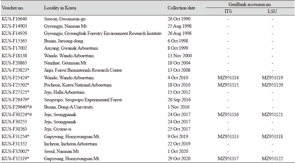
여름포자 세대의 형태적 특징을 파악하기 위하여 2008년 이후에 채집된 총 10점의 시료를 검경하였다(Table 1). 여름포자퇴(uredinia)와 여름포자(urediniospore)의 형태적 특징을 파악하고 크기를 측정하기 위해 해부현미경(SZX12, Olympus, Tokyo, Japan)과 명시야광학현미경(BX51, Olympus, Tokyo, Japan)을 사용하였고, 현미경사진은 AxioCam MRc5 카메라(Zeiss AX10, Carl Zeiss, Germany)를 사용하여 촬영하였다. 여름포자의 외부 형태와 미세구조는 주사전자현미경(SUPRA 55VP, Carl Zeiss, Germany)을 이용하여 관찰 및 촬영하였다. 겨울포자 세대는 2점의 시료에서만 관찰되었으며(Table 1), 때죽나무 잎에서 노화된 병반을 동결마이크로톰(Leica CM 3050 S, Leica Biosystems, Wetzlar, Germany)을 이용하여 10 μm 두께로 잘라 Lactophenol cotton blue로 염색한 후 관찰하였다.
병 초기에는 잎에 엽맥으로 제한된 다각형의 퇴록 병반이 형성되었고, 점차 병반이 합쳐지고 커지면서 후기에는 갈색으로 변했다(Fig. 1B and 1C). 녹병이 심한 경우에는 조기낙엽을 일으켰다(Fig. 1A). 여름포자퇴는 대부분 잎 뒷면에 형성되었으며, 군데군데 흩어져 있었고(Fig. 1D), 노란색으로 다소 돌출되었으며, 대부분 원형으로 직경은 0.1-0.2 mm였고, 바깥쪽 표피조직은 정단부에서부터 불규칙하게 터지면서 여름포자가 드러났다(Fig. 1F). 여름포자(n=30)는 계란형 내지 타원형으로 크기는 20-29 × 13.5-19 μm (평균 24.04 × 16.05 μm)였으며, 세포벽 두께는 0.9-1.5 μm였고, 표면에 원뿔형의 미세한 돌기가 전체적으로 분포하였다(Fig. 1G and 1H). 겨울포자(teliospore)가 형성된 부위는 엽맥으로 경계를 이룬 갈색 내지 흑갈색 병반으로 보였다(Fig. 1I). 겨울포자(n=30)는 주로 잎 뒷면의 표피층 아래에서 관찰되었고, 2-4개의 세포로 이루어졌으며 각각의 세포의 크기는 9-21 × 25-44 μm (평균 14.46 × 36.66 μm)였다(Fig. 1J and 1K). 이러한 형태적 특징은 겨울포자의 길이가 다소 긴 것을 제외하고는(일본 기록 10-24 × 18-30 μm) 일본의 쪽동백나무(Styrax obassia)에서 보고된 Pucciniastrum styracinum Hiratsuka의 기재와 일치하였다[9].
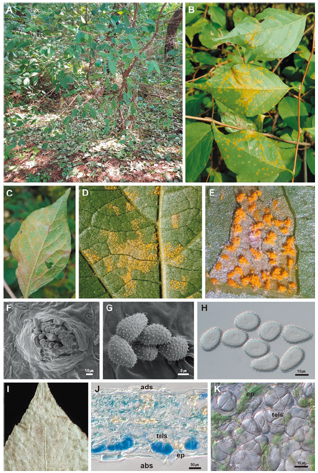
Fig. 1. Pucciniastrum styracinum occurring on Styrax japonicus. A, early defoliation of an affected tree in late September, 2020. B, symptoms on the lower leaf surface of affected leaves in the early stage. C, symptoms on the lower leaf surface in the advanced stage. D and E, uredinia formed on the lower leaf surface. F and G. Uredium (F) and urediniospores (G) observed under a scanning electron microscope. H, urediniospores focused on outline under a DIC microscope. I, symptoms on the lower leaf surface with the telial stage. J, vertical section of teliospores on the leaf epidermis on the abaxial side. K, cross section of teliospores seen from the leaf epidermis. abs, abaxial side; ep, epidermis of the leaf; tells, teliospores.
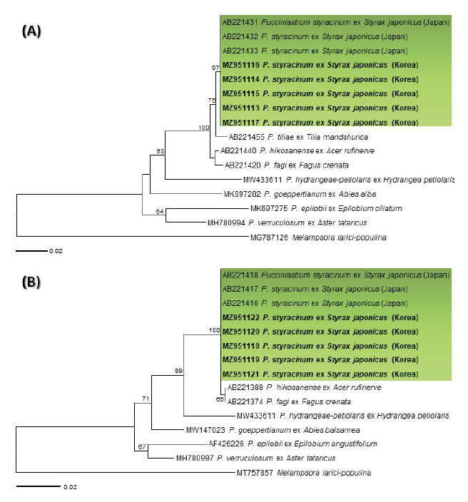
때죽나무 녹병균의 동정을 위해서, internal transcribed spacer (ITS) 및 large subunit (LSU) rDNA 영역의 염기서열을 이용하여 분자계통분석을 수행하였다. 동일한 10개의 건조시료에서 채취한 여름포자에서 MagListoTM 5M Plant Genomic DNA Extraction Kit (Bioneer, Daejeon, Korea)를 이용하여 genomic DNA를 추출하였고, ITS5u/ITS4rust [10] 및 LRust1R/LRust3 [11] 프라이머 조합을 사용하여 ITS와 LSU 영역을 각각 증폭하였다. PCR 조건은 95℃에서 4분(initial denaturation), 95℃에서 40초(denaturation), 54℃에서 1분(annealing), 72℃에서 1분 20초(elongation), 72℃에서 7분(final elongation)이며, denaturation에서 elongation단계까지 36회 반복하여 진행하였다. 그 중 5개 시료의 genomic DNA에서 증폭된 PCR 산물을 전기영동하여 확인한 후, AccuPrep® PCR/Gel DNA Purification Kit (Bioneer, Daejeon, Korea)를 이용하여 정제하여 마크로젠(Macrogen, Seoul, Korea)에 염기서열 분석을 의뢰하였으며, PCR 증폭에 쓰인 동일한 프라이머로 염기서열분석을 진행하였다. 분석된 염기서열은 DNAStar software package 7.1 (Lasergene, Madison, WI, USA)를 이용하여 편집 및 정리하였으며, NCBI (National Center for Biotechnology Information)의 GenBank에 등록하였다(Table 1). BLASTn을 이용하여 GenBank에 등록된 염기서열과 비교한 결과, ITS 영역 부위의 염기서열은 GenBank에 등록된 일본의 때죽나무 녹병균 P. styracinum의 염기서열(AB221431-AB221433)과 100% 일치하였으며, LSU 영역의 염기서열도 일본 때죽나무 녹병균(AB221416-AB221418)과 100% 일치하였다. 이들의 계통학적 유연관계를 분석하기 위해서 MEGA 7.0 program [12]의 maximum likelihood 방법으로 ITS 및 LSU 염기서열에 대한 계통수를 각각 작성하였다(Fig. 2). 염기서열의 유전적 거리는 Tamura-Nei model로 계산하였고, bootstrap 분석은 1,000 반복으로 수행하였다. ITS 및 LSU 염기서열의 계통수(Fig. 2A and 2B)에서 한국의 때죽나무 녹병균 시료는 일본의 때죽나무 녹병균 P. styracinum과 각각 97, 100%의 높은 bootstrap 신뢰도지수를 가지고 하나의 그룹을 형성하였다.
때죽나무는 우리나라와 중국, 일본, 대만이 원산이지만 재래종이 도입되거나 품종이 개발되어 캐나다, 미국, 영국, 네덜란드에도 분포하고 있다[3]. Pucciniastrum styracinum에 의한 때죽나무 녹병은 일본과 우리나라에서 보고되었으나, 녹병균에 대한 상세한 정보는 P. styracinum을 신종으로 최초 보고한 1898년 일본의 기록이 유일하다[13,14]. 본 연구에서는 우리나라에서 발생하는 때죽나무 녹병균의 형태적 특징 및 분자 분석의 결과를 종합하여 녹병균을 P. styracinum으로 동정하였고, 때죽나무에서 발생하는 여름포자 세대와 겨울포자 세대의 균학적 특성과 형태적 측정치 및 분자적 특성에 대한 정보를 제공하였다.
우리나라에서 때죽나무 녹병균은 1967년 임업시험장(현, 국립산림과학원)의 시험연구보고서(상편)에서 처음 기록되었다[8]. 이에 앞서 Hiratsuka는 일제강점시대 한반도의 녹병균을 비교적 상세하게 기록하였으나 지금은 비교적 흔히 발견할 수 있는 때죽나무 녹병균에 대한 기록은 당시에 없었다[15-19]. 또한 P. styracinum이 녹포자세대를 형성하는 중간기주로 전나무속의 Abies mayriana, A. sachalinensis var. mayriana, Abies sp.가 일본과 대만에서 보고되어 있지만 국내에 분포하는 전나무류에서는 Pucciniastrum속의 녹병균이 아직 기록되거나 발견된 적이 없다[7,13]. 따라서 1960년대 이후 국내 P. styracinum의 발생 원인을 밝히기 위해서는 Pucciniastrum속 대부분 녹병균의 중간기주인 전나무류의 분포 및 보급과 P. styracinum을 포함한 Pucciniastrum 녹병균의 지리적 분포와의 관계를 종합적으로 고찰할 필요가 있다.
Pucciniastrum styracinum에 의한 녹병은 일본과 중국, 대만에서 때죽나무, 쪽동백나무, 좀쪽동백나무(S. shiraianus), 대만때죽나무(S. formosanus), S. japonicus var. kotoensis, 일본나래쪽동백(Pterostyrax corymbosum), 나래쪽동백(P. hispidus)에서 보고되어 있으므로 국내 자생하는 쪽동백나무, 좀쪽동백나무와 재배식물인 일본나래쪽동백과 나래쪽동백도 관심있게 살펴볼 필요가 있다[13].
적요
때죽나무는 때죽나무과에 속하는 낙엽 소교목으로 우리나라와 중국, 일본, 대만이 원산이나 캐나다, 미국, 영국 등에도 도입되어 분포하고 있다. 1990년 이후 전국에 분포하는 때죽나무에서 녹병균이 지속적으로 발견되었다. 한국의 때죽나무 녹병균의 형태적 특성은 일본의 쪽동백나무에서 보고된 Pucciniastrum styracinum의 형태적 특성과 가장 유사하였으며 internal transcribed spacer (ITS) 및 28S large subunit (LSU) rDNA의 염기서열의 분자계통학적 분석을 통해 동정을 확인하였다. 본 연구에서는 한국의 때죽나무에서 발견한 녹병균 P. styracinum의 존재를 재확인하였고, 균학적 특성과 형태적 측정치 및 분자적 특성에 대한 정보를 제공하였다.


