서 론
내생균(endophytic fungi)은 식물에 공생하는 균류로, 숙주식물의 조직 내에서 서식하며 병원성을 띠지 않는 균류이다[1]. 내생균은 초본, 혹은 목본 식물체의 조직 내에서 독성 알칼로이드를 합성하여 포식자로부터의 보호 기작을 제공하기도 하고[2], 수분 스트레스[3], 식물 병원균 등에 대한 저항을 제공하기도 한다[4]. 본 연구에서는 제주도 한라산에 서식하는 구상나무(Abies koreana) 내생균의 다양성을 조사하던 중 구상나무의 침엽에서 두 종의 국내 미기록종을 분리하였으며, 그 특징을 보고하고자 한다.
재료 및 방법
한라산의 해발 1,800 m 주변에 서식하는 구상나무의 잎을 채취하였으며, 병증이 없는 건강한 침엽을 선별한 후 실험실로 운반하였다. 증류수로 세척된 침엽을 30%의 H2O2로 30초간 표면 살균한 뒤, potato dextrose agar (PDA) 배지에 4조각씩 치상하였다. 25°C의 암소에서 3~7일간 배양하면서 균사가 뻗어 나오면 새로운 PDA 배지에 계대하여 순수 분리하였고, 확보된 균주를 다시 PDA 배지와 malt extract agar (MEA) 배지에 삼점 계대하여 7일간 동일한 조건으로 배양한 뒤 형태적 특징을 관찰하였다(Fig. 1). 염기서열 분석을 위하여 DNeasy Plant mini kit (Qiagen, Germantown, MD, USA)의 protocol에 따라 균사에서 genomic DNA를 추출한 뒤 균 특이적인 프라이머인 ITS1F와 ITS4를 이용하여 internal transcribed spacer (ITS) 지역을 증폭하였고[5], 프라이머 LR0R과 LR16을 이용하여 rDNA의 large subunit (LSU) 지역을 증폭하였다[6]. Annealing 온도는 ITS 지역은 50°C, LSU rDNA 지역은 44°C로 설정하여 수행하였으며, PCR 산물은 1.5% agarose gel에서 22분간 전기영동을 실시하였고, 예상되는 크기의 DNA band를 확인한 후 염기서열 분석을 의뢰하였다(SolGent, Daejeon, Korea). 분석된 염기서열은 NCBI 상에서 BLAST를 이용하여 유사도를 확인한 후 각 종들 간의 계통 분석을 통한 유연 관계를 확인하기 위해 MEGA6 [7]를 이용하여, ITS 지역의 DNA 염기서열과 LSU 지역의 염기서열을 combined-sequences로 align하여 neighbor- joinning 방법으로 phylogenetic tree를 작성하였다(Figs. 2, 3). 분리된 균주는 국립생물자원관(NIBR)에 기탁하였다.
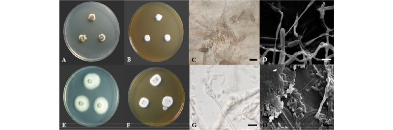
Fig. 1. Colonies of Coleophoma parafusiformis 17E082 grown on PDA (A) and MEA (B), conidiophore (C, optical microscope image; D, scanning electron microscope image). Colonies of Coniochaeta ligniaria 16H328 grown on PDA (E) and MEA (F), conidiophore and conidia (G, optical microscope image; H, scanning electron microscope image). PDA, potato dextrose agar; MEA, malt extract agar (scale bars = 10 µm).
결과 및 고찰
Coleophoma parafusiformis Crous, Fungal Biol 120: 1405 (2016)
PDA 배지에서 7일간 배양된 균총의 직경은 12~13 mm 정도이며, 균총의 색은 앞면은 밝은 크림색이고 뒷면은 적갈색을 띤다. 고도는 배지에 납작하게 붙어 있고, 가장자리는 불규칙한 방사형으로 뻗어 있다(Fig. 1A). MEA 배지에서 7일간 배양된 균총의 직경은 10~12 mm 정도이며, 균총의 색은 앞면은 전체적으로 흰색을 띠고 뒷면은 중앙부에서 적갈색을, 가장자리에서 흰색을 띤다. 균총의 고도는 중앙부에서 볼록 융기되어 있으며, 가장자리의 형태는 얇은 균사들이 방사형으로 뻗어 나가는 형태이며 간혹 불규칙하게 파여 있는 가장자리의 형태를 확인할 수 있다(Fig. 1B). 분생자경의 형태는 부드러운 타원형 혹은 원통형으로 균사 끝에서 솟아오른 형태이고, 격벽이 없는 것에서부터 2~3장의 격벽을 가진 것까지 다양한 형태가 존재한다. 분생자경의 크기는 (22.7~28.4) × (8.6~11.1) µm 정도이며, 색은 짙은 갈색 혹은 검은색이다(Fig. 1C).
Specimen examined: Mt. Hallasan, Jeju-do, Korea, N 33°21'47.9", E 126°32'06.1", April 18, 2017, isolated from leaves of Abies koreana, strain 17E082, NIBRFG0000502338, GenBank no. MG905602.
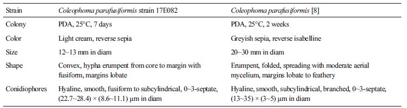
Note: C. parafusiformis는 2016년 Crous [8]에 의해 신종으로 명명된 종이다. 균사의 생장 방향 측면에 방추형(fusiform)의 길쭉한 분생자경(conidiophore)을 갖는 것이 특징이며, 동유럽 라트비아의 진달래속(Rhododendron) 식물의 잎에서 내생균으로 분리되었다[8]. 균총 및 분생포자의 형태적 특징은 대체로 원 기재문과 일치하였으며(Table 1), ITS 지역과 LSU 지역의 염기서열의 분석 결과 ITS 지역의 DNA 염기서열은 C. parafusiformis KU728494.1과 99%의 일치도를 보였고, LSU 지역의 염기서열은 C. parafusiformis KU728534.1과 98%의 일치도를 보였으며, 모두 같은 계통을 형성하고 있었다(Fig. 2).
Table 1. Morphological characteristics of Coleophoma parafusiformis strain isolated from this study
|
|
|
PDA, potato dextrose agar. |
|
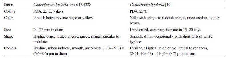
Table 2. Morphological characteristics of Coniochaeta ligniaria strain isolated from this study
|
|
|
PDA, potato dextrose agar. |
|
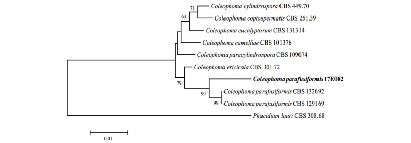
Fig. 2. Neighbor-joining phylogenetic tree based on a combined alignment of both internal transcribed spacer (ITS), large subunit (LSU) sequences. Phacidium lauri was used as an outgroup. Numbers on branches indicate bootstrap values (1,000 replicates). Fungal strain isolated in this study are in bold.
Coniochaeta ligniaria (Grev.) Massee, Grevillea 16: 37 (1887)
PDA 배지에서 7일간 배양된 균총의 직경은 20~23 mm 정도이며, 균총의 색은 앞면은 분홍빛이 감도는 옅은 베이지색을 띠고, 뒷면은 베이지색 혹은 노란색이다. 중앙부에는 균사가 밀집하여 살짝 융기한 형태이며, 가장자리는 둥근 형태이다(Fig. 1E). MEA 배지에서 7일간 배양된 균총의 직경은 18~20 mm 정도이며, 균총 앞면의 색은 중앙부에서 연분홍색을 띠고 가장자리는 베이지색이고, 균총 뒷면의 색은 흰색에 가깝다. 균총의 고도는 배지에 납작하게 붙어 있으며, 가장자리는 방사형으로 조밀한 균사가 뻗어 나가는 형태이다(Fig. 1F). 균사 끝이 비대해져서 형성된 분생자경에서 원통형의 분생자가 형성되며, 분생자는 투명한 유리질이고 크기는 (17.4~22.3) × (6.6~8.6) µm 정도이다(Table 2, Fig. 1G).
Specimen examined: Mt. Hallasan, Jeju-do, Korea, N 33°22'22.1", E 126°33'03.1", August 23, 2016, isolated from leaves of Abies koreana, strain 16H328, NIBRFG0000502332, GenBank no. MG905601.
Note: C. ligniaria는 1887년 Massee에 의해 최초로 기록된 종이다[9]. 본 종은 자낭균류에 속하는 균 중 perithecium을 갖는 Pyrenomycetes에 속하며, 일반적으로 목본식물에 서식하는 내생균이다[10]. 항진균성을 갖는 지방산 화합물을 생성하여 식물병의 원인이 되는 진균에 대한 내성을 제공하는 것으로 연구된 바 있다[11]. ITS 지역과 LSU 지역의 분자적 분석 결과 ITS 지역의 DNA 염기서열이 C. ligniaria AY198390.1과 99%의 일치도를 보였고, LSU 지역의 염기서열은 C. ligniaria AY198388.1과 100%의 일치도를 보였으며, 모두 같은 계통을 형성하였다(Fig. 3).
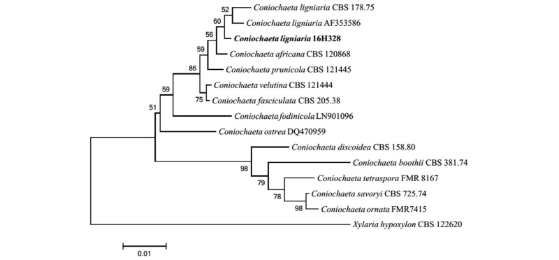
Fig. 3. Neighbor-joining phylogenetic tree based on a combined alignment of both internal transcribed spacer (ITS), large subunit (LSU) sequences. Xylaria hypoxylon was used as an outgroup. Numbers on branches indicate bootstrap values (1,000 replicates). Fungal strain isolated in this study are in bold.
