서론
우리나라에서 복숭아나무(복사나무, Prunus persica var. persica)의 흰가루병균으로 Phyllactinia corylea P. Karst., Podosphaera pannosa (Wallr.) de Bary, Podosphaera tridactyla (Wallr.) de Bary의 3종이 기록되어 있다[1]. P. corylea는 1928년에 기록된[2] 이후에 한국에서 추가로 확인된 적이 없다[3, 4]. P. pannosa는 1991년에 Dong 등[5]에 의해 복숭아 과실에서 처음으로 보고되었으며, 농촌진흥청 농업기술연구소에 의해 추가로 기록되었다[3]. P. tridactyla는 1958년에 Park [6]에 의해 복숭아나무에서 처음으로 보고되었으며, 농촌진흥청 농업기술연구소에 의해 잎에 발생되는 것으로 추가 기록되었다[3].
그 중, 복숭아 과실을 침해하는 흰가루병균 P. pannosa의 동정은 무성세대의 형태적 특징에 의거하였으며[5], 실질적으로 P. tridactyla의 무성세대와 형태적으로 구분하기 어려우므로[7] 재확인이 필요하다는 의견도 있었다[4]. 그러나, 유럽[8], 미국[9], 일본[10]에서 복숭아나무 흰가루병균으로 P. pannosa를 보고하면서 발병 부위에 과실을 포함시키거나 병징이 나타난 복숭아 과실 사진을 제시하기도 하였다. 따라서 복숭아 과실의 흰가루병균은 P. pannosa로 알려져 왔다.
한편, 유럽에서는 Jankovics 등[11]이 복숭아 과실의 흰가루병균으로 P. leucotricha를 발표함으로써 복숭아 과실의 흰가루병균에 대한 정체성에 의문점이 제기되었다. 따라서 본 연구는 한국에서 기록된 복숭아 과실의 흰가루병균의 정체성을 확실하게 밝힘으로써 Prunus 기주에서의 Podosphaera 흰가루병균과의 관계를 이해하는 데 일조하고자 수행하였다.
시료 채집
복숭아 과실에 발생한 흰가루병균을 총 4회 채집하였다. 그 중 3점을 고려대학교 진균표본실(KUS)에 보존하였다. 소장된 시료의 내역은 다음과 같다. KUS-F28653 (19 Jun 2015, 강원도 춘천시 신북읍 발산리, 품종 ‘황도’), F29798 (14 Jun 2017, 강원도 춘천시 신북읍 유포리, 품종 ‘백향’), F29842 (4 Jul 2017, 세종특별자치시 조치원읍 번암리, 품종 ‘미홍’) 등 3점이었다. 그 외에, 2011년 8월 5일 경상북도 김천시의 복숭아 과수원에서 종이 봉지를 씌워 키운 ‘황도’에서 수확 당시에 흰가루병 병반이 뚜렷한 과실이 발견되었다. 이 병반은 현미경 관찰을 통해 흰가루병균의 분생포자경과 분생포자를 관찰하여 P. pannosa로 동정하였으나 표본으로 제작하기 전에 과실이 부패함으로써 표본으로 보존하지는 못했다.
병징학적 특징
복숭아 과실의 흰가루병균은 과실 표면에 흰색의 집락으로 발견되었다. 유과기에 흰가루병균의 집락은 뚜렷하면서 전형적인 흰가루(powdery mildew) 증상을 나타냈다(Fig. 1A). 그러나, 일찍 감염되거나 집락이 노화될수록 과실 표면이 변색되어 전형적인 녹얼룩점(rusty spot) 증상을 나타냈다(Fig. 1D, 1E). 이러한 병징은 대부분 어린 과실에서 나타났으나, 수확기의 성숙한 과실에서도 새로운 흰가루병균 집락이 나타나기도 하였다(Fig. 1B, 1C).
흰가루 증상에서는 병반의 안쪽이나 가장자리에서 모두 분생포자경과 분생포자가 뚜렷하였다. 그러나 녹얼룩점 증상에서는 병반의 안쪽에서는 분생포자경과 분생포자가 노화되어 형태적 특징의 검경에는 적합하지 않았으며, 병반의 가장자리에서는 일부 분생포자경과 분생포자를 확인할 수 있었다. 따라서 흰가루 증상과 녹얼룩점 증상이 감염시기나 품종에 따른 특성으로 보여졌으며 흰가루병균은 형태적으로 차이가 없었다.
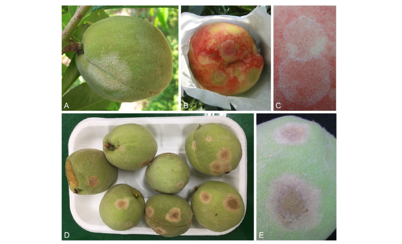
Fig. 1. Symptoms on peach (Prunus persica var. persica) fruits associated with a powdery mildew fungus, Podosphaera pannosa. A, Powdery mildew symptom on a young fruit ‘Hwangdo’; B, Powdery mildew symptoms on a mature fruit ‘Hwangdo’; C, Close-up of powdery mildew symptoms on a mature fruit ‘Hwangdo’; D, Rusty spot symptoms on young fruits ‘Mihong’; E, Close-up of rusty spot symptoms on a young fruit ‘Mihong’.
형태학적 특징
복숭아 과실에 발생한 흰가루병균의 무성세대를 검경하였다. 분류학적 특성을 파악하고 크기를 측정하기 위해서 명시야광학현미경(BX51; Olympus, Tokyo, Japan)을 사용하였으며, 현미경 사진은 미분간섭현미경(Axio Imager; Carl Zeiss, Oberkochen, Germany)을 이용하였다. 무성세대의 분류학적 특징 파악 및 현미경 사진 촬영은 신선 시료를 사용하여 수행하였으며, 증류수를 검경액으로 사용하여 수행하였다.
균사는 과실의 표면에 존재하며, 대부분 파상이며, 때로는 결절을 형성하였다. 균사 부착기는 발달이 미약하며, 유두상이며, 단생하였다(Fig. 2A, 2B). 분생포자경은 표생균사의 윗부분으로부터 발달하며, 110~170 × 8~10 µm이며, 3~8개의 세포로 구성되며, 분생포자를 연쇄상으로 형성하였다(Fig. 1E). 분생포자경의 기부세포는 아래쪽이 곧고, 길이는 40~70 µm였다(Fig. 2C~2E). 분생포자는 무색의 단세포이며, 뚜렷한 피브로신체를 가지며, 타원형 내지 달걀형이며, 크기는 25~38 × 12~16 µm였다(Fig. 1F, 1G). 유성세대의 흔적은 발견되지 않았다. 이러한 균학적 특징은 앞선 연구에서 보고된 Podosphaera pannosa와 일치하였다[12].
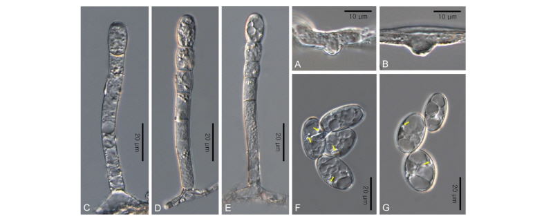
Fig. 2. Phylogenetic relationship between two Podosphaera pannosa isolates ex Prunus persica var. persica and some reference isolates retrieved from GenBank, inferred by maximum likelihood method using the internal transcribed spacer regions. Bootstrap values based on 1,000 replications are indicated above the branches. The scale bar represents 0.02 nucleotide substitutions per site. The Korean isolates presented in this study are indicated in bold.
분자계통학적 분석
복숭아 과실을 감염하는 흰가루병균 보존시료 중에서 KUS-F28653 및 F29842를 선정하여 internal transcribed spacer (ITS) 영역의 염기서열을 분석하였다. KUS-F29798 시료에서는 rDNA 추출에 실패하여 본 분자계통학적 분석에는 포함시키지 못했다. Takamatsu 등[13]의 방법에 따라 시료에서 채취한 균사와 분생포자로부터 rDNA를 추출하였고, ITS5 [14]와 P3 [15] 프라이머를 사용하여 polymerase chain reaction (PCR)으로 증폭시켰다. 증폭된 PCR 산물을 전기영동을 통하여 확인한 후에 QIAquick PCR Purification kit (Qiagen, Germantown, MD, USA)로 정제하였다. 이 염기서열을 DNASTAR Lasergene computer package 5.05 (DNASTAR, Madison, WI, USA)를 이용하여 정리한 후, GenBank에 등록하여 기탁번호를 부여 받았다(accession nos. MH252368, MH252369). GenBank BLAST를 이용하여 ITS 영역의 염기서열을 비교한 결과, 한국에서 채집된 복숭아 과실의 흰가루병균은 장미류(Rosa spp.) 흰가루병균인 P. pannosa와 99% 이상 일치하였다. 이 결과를 바탕으로 MEGA7 프로그램[16]을 이용하여 maximum likelihood 방법으로 계통수를 작성하였다. 그 결과 복숭아 과실의 흰가루병균은 P. pannosa의 계통군에 속하였다(Fig. 2). 따라서 한국의 복숭아 과실을 감염하는 병원균은 P. pannosa임을 분자적으로 처음 확인하였으며, 흰가루 증상 및 녹얼룩점 증상 모두 P. pannosa에 의해 발생됨을 확인하였다.
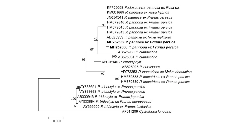
Fig. 3. Phylogenetic relationship between two Podosphaera pannosa isolates ex Prunus persica var. persica and some reference isolates retrieved from GenBank, inferred by maximum likelihood method using the internal transcribed spacer regions. Bootstrap values based on 1,000 replications are indicated above the branches. The scale bar represents 0.02 nucleotide substitutions per site. The Korean isolates presented in this study are indicated in bold.
고 찰
지금까지 유럽에서는 과수원에서 재배하는 복숭아나무의 잎을 감염하는 대표적인 흰가루병균으로 Podosphaera pannosa와 P. tridactyla가 알려져 있다[10]. 반면, 미국에서는 복숭아나무의 과실 흰가루병균으로 P. pannosa [17]와 P. leucotricha [9]가 알려졌다. 최근에 세르비아와 프랑스의 과수원에서 채집한 10점의 시료를 바탕으로 연구한 Jankovics 등[11]의 결과에 따르면, 복숭아 과실에서는 녹얼룩점 증상이 나타나며 P. leucotricha가 관여하고, 천도 과실에서는 흰가루 증상이 나타나며 P. pannosa가 관여하는 것으로 보고했다.
한편, 장미과(Rosaceae) 식물에 발생하는 Podosphaera속 흰가루병균에 대한 균학적 연구를 통하여 Prunus속 식물의 흰가루병균은 대부분 P. tridactyla이지만 일부는 P. clandestina (Wallr.) Lév.로 동정하였다[18]. 더구나 P. tridactyla는 단계통군이 아니라 유전적 다양성이 상당히 넓은 복합종으로 여겨진다고 보고하였다[18]. 이들 연구에서는 흰가루병균이 잎에서 유래한 것인지 과실에서 유래한 것인지를 밝히지 않았으나, 특별한 언급이 없는 것으로 보아 모두 잎에서 유래한 것으로 여겨진다.
따라서, 전 세계적으로 지금까지 복숭아나무에서 기록된 흰가루병균은 P. clandestina, P. leucotricha, P. pannosa, P. tridactyla 등 4종이다. 이 중 P. clandestina가 복숭아 과실에서 기록되었는지 여부는 불확실하지만, 나머지 3종의 흰가루병균이 복숭아 과실에서 기록된 것은 확실하다. 이 중에서 복숭아 과실에서의 존재를 분자계통학적으로 확인한 것은 P. leuchotricha가 유일하다. 우리나라를 비롯한 일부 국가에서 복숭아 과실 흰가루병균으로 기록된 P. pannosa를 본 연구를 통하여 분자계통학적으로 처음 확인하였다.
