서론
쑥부쟁이(Aster yomena)는 국화과에 속하는 다년생 초본으로 한국을 비롯한 일본, 중국, 시베리아에 분포하며 근경이나 종자로 번식한다. 민간에서는 전초는 ‘마란’, 지상부는 ‘백상국'으로 불리며, 한방에서 소풍, 청열, 해독, 거담진해, 이뇨, 보익, 해소 등의 약으로 이용되어 왔다[1]. 그리고 어린순을 데쳐먹거나 기름에 볶아서 먹기도 하는 산채나물로 이용되어 왔으며, 식욕을 촉진시키는 것으로 알려져 있다[2]. 최근 산채에 대한 기능성 건강식품으로 관심이 높아지면서 각종 산채에 대한 항산화, 항암, 항염증 등 생리활성 연구가 진행되고 있으며, 쑥부쟁이 에탄올 추출물이 간세포 내 지방축적 유전자 발현 억제와 지방 축적 억제 효과가 있다고 보고한 바 있다[3].
쑥부쟁이 균핵병 발생
쑥부쟁이 균핵병은 2016년에 전남 구례에서 쑥부쟁이를 재배하는 비닐하우스 포장과 노지 포장에서 발생하였으며(Fig. 1F, 1G), 포장에서 토양 수분이 많은 곳부터 발생하는데 9월부터 발병이 시작하여 다음 해 5월까지 발생하였고, 발생한 피해 포기가 20~80%까지 발생하여 심지어 쑥부쟁이 재배를 포기할 정도로 쑥부쟁이 생산에 큰 피해를 주는 병이다(Table 1). 쑥부쟁이 균핵병의 병징은 쑥부쟁이 식물체 캐노피 내에 쑥부쟁이 줄기가 물러지고 균핵병균의 흰색 균사로 실가닥으로 뒤덮여 있으며(Fig. 1A), 진전되면 식물체가 갈변되어 썩어 물러지면서 썩는다(Fig.1B, 1C, 1D). 일부 병든 식물체는 줄기속에서 소형 흑색 균핵을 형성하기도 한다(Fig. 1E).
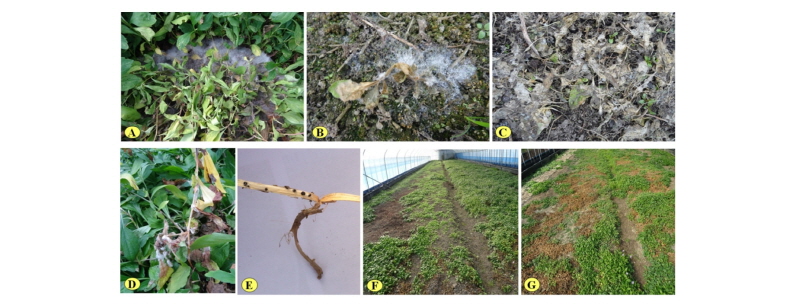
Fig. 1. Symptoms and damages of Aster yomena Sclerotinia rot caused by Sclerotinia minor in Gurye. A~B, diseased plant and soil surface covered with white and fluffy mycelia of S. minor and watery soft lesions of plants on November in 2016; C, dried diseased plants on soil; D, diseased stems of a plant with white and fluffy mycelia of S. minor; E, black small sclerotia of S. minor inside diseased stem; F, damage in greenhouse; G, damage in open field.
|
Table 1. Occurrence of Sclerotinia rot of Aster yomena by Sclerotinia minor in Gurye on April in 2016 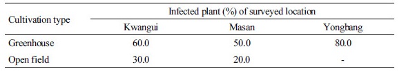
|
시료 채취 및 균핵병균 분리
2016년에 전남 구례군의 쑥부쟁이 재배지역별로 비닐하우스 포장과 노지 포장에서 병 걸린 식물체를 채집하여 병 걸린 경계부위 조직을 잘라서 1% 차아염소산나트륨용액(NaOCl)에 20초간 침지한 후 멸균수로 3회 수세하여 건조하였다. 건조한 이병조직을 물한천배지(water agar)에 치상하여 20°C에 배양하면서 자란 단균사를 떼어 내어 potato dextrose agar (PDA; Difco, Detroit, MI, USA) 사면 배지에 치상하여 20°C에서 14일간 배양한 분리균주를 10°C 항온기에 보관하면서 실험에 사용하였다.
분리균주의 DNA 추출 및 염기서열 분석
분리균주의 염기서열 분석을 위하여 genomic DNA를 추출하였다. 분리한 6개 균주(SGB31, SGB41, SGC33, SGC35, SGY31, SGY34)를 PDA배지에 각각 접종하고, 20°C에서 7~10일간 배양한 후, PDA 배지에 형성된 균핵을 수거하여 동결건조하고 마쇄하였다. CTAB- phenol/chloroform을 이용한 Choi 등[4]의 방법으로 genomic DNA를 추출하고, ‑20°C에 보존하면서 실험에 사용하였다. Internal transcribed spacer (ITS) rDNA 염기서열 분석을 위하여 ITS1F/ITS4 프라이머를 사용하여 polymerase chain reaction (PCR) 증폭을 수행하였다[5]. PCR 반응액은 100 ng/µL의 template DNA를 포함하여 10× Taq buffer, 2 mM dNTPs, 10 pmole/µL의 양방향 primer쌍, 0.5 unit의 Taq DNA polymerase (Takara Bio, Kusatsu, Japan)를 총량 50 µL로 제조하였다. PCR 증폭은 Bio-Rad tetrad 2 thermal cycler (Bio-Rad, Hercules, CA, USA)를 이용하여 94°C 30초, 52°C 30초, 72°C 90초를 35회 반복하였고, 최종적으로 72°C에서 7분간 post extension을 실시한 후, 1% agarose gel에 전기영동하여 band를 확인하였다. 증폭된 band는 Wizard SV Gel & PCR Clean-Up System kit (Promega, San Luis Obispo, CA, USA)를 사용하여 정제하고, direct sequencing으로 염기서열을 분석하였다. 분석된 염기서열은 Clustal W 소프트웨어[6]를 이용하여 정렬하였고, 계통수는 MEGA 6.0 프로그램을 이용하여 neighbor-joining법에 의해 작성하였으며, out-group으로는 Botrytis cinerea CBS:131.28 균주(KF859918)를 사용하였다.
MEGA6.0 프로그램을 이용하여 분자유전학적인 계통도를 작성하고 분리균주의 분류학적 위치를 확인하였다. 작성한 계통도에서 분리균주는 KF859929 Sclerotinia minor CBS:339.39 KF859930와 Sclerotinia minor DAOM:191806 균주와 높은 상동성(100%)을 보였다(Fig. 2). 계통도의 신뢰성을 확보하기 위한 Bootstrap 값도 94%로 비교적 높은 값을 보였다.
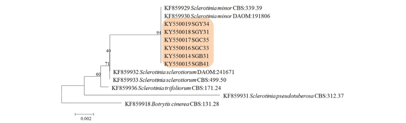
Fig. 2. Phylogenetic relationship between Sclerotinia minor and some reference isolates retrieved from GenBank, inferred by neighbor-joining method using the internal transcribed spacer rDNA region. Bootstrap values based on 1,000 replications are indicated above the branches and the scale bar represents 0.002 nucleotide substitutions per site.
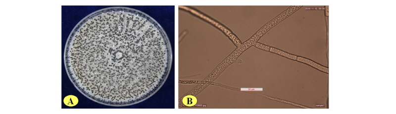
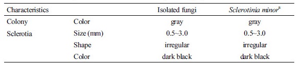
분리균의 형태적 특성
분리균을 20°C에서 14일간 PDA에서 배양한 균총은 잿빛이었으며, 배지 표면에 불규칙 소형 흑색 균핵을 형성하였으며 그 크기는 0.5~3.0 mm이었다(Fig. 3A, Table 2), 분리균을 물한천배지에서 20°C에서 3일간 배양한 균사를 형광현미경 (Leica DM2500, Leica Microsystems, Wetzlar, Germany)으로 관찰하였다(Fig. 3B). 이들 분리균주는 Damon [7]의 균주와 비교하여 형태적으로 유사하였으며, 분자적 염기서열을 분석한 결과 Sclerotinia monor로 동정하였다.
또한 쑥부쟁이 균핵병균(Sclerotinia minor) SGC33 (KACC 48270), SGY31 (KACC 48271), SGB31 (KACC 48282) 균주는 농촌진흥청 농업미생물은행(KACC)에 기탁하였다.
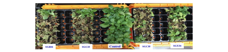
분리균의 병원성 검정
50공 트레이포트에 바로크상토(Seoul Bio, Eumseong, Korea)를 담고, 쑥부쟁이 종자를 파종하여 4주간 생육한 식물체에 분리균주(SGB41, SGC33, SGC35, SGY34)를 PDA 배지에서 22°C에서 6일간 배양한 균총을 직경 5 mm 코르코보러로 떼어내어 식물체의 지제부 줄기에 부착하여 균핵병 발생을 조사한 결과, 접종 2일 후부터 발병하기 시작하였으며, SGB41, SGC33, SGC35, SGY34 균주 순으로 병 발생이 심하였으며(Fig. 4), 병이 발생한 쑥부쟁이 줄기에서 균을 재분리하여 접종한 균과 동일한 균을 확인하였다.