서론
진균은 자연 생태계에서 유기물의 분해자로서 커다란 생태적 기능을 수행하는 미생물로서 종 다양성이 매우 높은 생물이다. 특히 나고야의정서 이후 진균의 종의 가치는 그 다양성과 더불어 유전자원으로서 매우 가치가 높은 실정이다. 국내에서도 이러한 국제적 추세와 더불어 생물종 발굴에 대한 노력으로 공기, 흙, 식물, 동물 등 다양한 환경에서 진균이 분리 및 동정되었다. 특히 최근 미기록 종에 대한 연구를 살펴보면 분리된 원천은 공기, 토양, 식물의 뿌리, 곤충 등으로 다양하였다[1-7]. 이렇듯 다양한 환경에서 미기록 종이 발견이 됨에 따라 아직 진균에 대한 연구가 되어 있지 않은 지역에서 진균 발굴에 대한 연구가 꾸준히 요구되고 있다.
연천군은 경기도의 최북단에 위치하고 있으며 겨울에는 시베리아 기단의 한랭 건조한 날씨가 지속되고 여름에는 북태평양 기단의 영향을 받아 고온 다습한 특징을 보이는 지역이다. 연천군은 마식령 산맥과 광주산맥에 의해 형성된 해발 200 m 이상의 연속된 산지들이 다른 지자체와 경계를 이루고 있으며, 자연생태환경의 보고인 비무장지대(DMZ)와 민간인통제선 구역의 약 50.1%를 차지하고 있다[8]. 정부는 연천군 DMZ 일원을 관광지로 개방하였는데 태풍전망대와 열쇠전망대가 있다. 기존에 보고된 연천군 지역에 대한 생물조사로는 물거미, 식물분포 등에 대한 연구가 있으나 아직 진균에 대한 조사연구는 보고되어 있지 않은 실정이다[9-11]. 이에 따라 본 연구는 자생진균 발굴의 일환으로 경기도 연천군의 DMZ 일원에 존재하는 진균의 다양성을 알아보고자 수행되었다. 이를 위해 2017년 5월에는 열쇠전망대 인근의 산에서 그리고 8월에는 태풍전망대 인근 임진강변에서 야생식물과 토양으로부터 시료를 각각 채집하였다. 채집된 시료로부터 진균을 동정하고 이중 다섯 종의 미기록 진균을 확인하였고 이들의 형태적, 분자적 특성에 대해서 보고하고자 한다.
재료 및 방법
시료 채집 및 순수분리
토양
토양 시료는 2017년 5월과 8월에 경기도 연천군 열쇠전망대 인근 마전리에 위치한 산과 태풍전망대 인근 중면 황진리 임진강변에서 각각 채집하였다. 토양 시료 차출은 토양 요거를 사용하여 표토로부터 10 cm 지점의 토양을 채취한 후 지퍼백에 담아 아이스쿨러에 보관하여 실험실로 운반하였다. 진균분리를 위해서는 멸균된 증류수 20 mL에 토양시료 1 g을 넣고 20분간 vortexing을 하여 고르게 섞어준 후 혼탁된 시료액을 암피실린이 첨가된 potato dextrose agar (PDA; Difco, Detroit, MI, USA)에 100 μL씩 평판도말 후 25℃에서 3일간 배양하면서 피어나는 균사를 순수분리 하였다[12].
식물체
경기도 연천군 열쇠전망대 인근 마전리에 위치한 산에서는 여뀌(Persicaria hydropiper), 붉나무(Rhus javanica), 족제비싸리(Amorpha fruticosa), 고비(Osmunda japonica), 개암나무(Corylus heterophylla var. thunbergii)의 잎(Fig. 1A)을 채집하였고 태풍전망대 인근 중면 황진리 임진강변에서는 사시나무(Populus davidiana Dode), 복숭아나무(Prunus persica), 은사시나무(Populus tomentiglandulosa), 달뿌리풀(Phragmites japonica), 물박달나무(Betula dahurica), 바랭이(Digitaria ciliaris)의 잎을 시료로 채집하였다(Fig. 1B). 채집된 시료는 시료에서 병반처럼 보이는 부위를 중심으로 해부현미경(SZ61 stereo microscope; Olympus, Tokyo, Japan)으로 관찰 후 부위의 일부를 생물안전작업대에서 멸균된 수술용 칼을 이용하여 1 ⅹ 1 cm 크기의 단편으로 절단하였다. 자른 절편시료는 2% 차아염소산나트륨 용액에 1분, 100% 에탄올에 1분, 멸균된 증류수에 1분씩 처리하였다가 멸균 필터페이퍼로 물기를 제거한 후 암피실린이 첨가된 PDA에 올린 다음 25℃에서 배양하면서 균사의 발생을 유도하였다[12, 13].
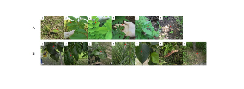
Fig. 1. Photos of soil and plant sources sampled in this study for the isolation of fungi in the vicinity of DMZ in Yeoncheon-gun. A row, Sampled in Majeon-ri; B row, Sampled in Hwangjin-ri; A1, Persicaria hydropiper; A2, Rhus javanica; A3, Amorpha fruticosa; A4, Osmunda japonica; A5, Corylus heterophylla var. thunbergii; A6, Soil; B1, Populus davidiana Dode; B2, Prunus persicaa; B3, Populus tomentiglandulosa; B4, Phragmites japonica; B5, Betula dahurica; B6, Digitaria ciliaris; B7, Soil; Red arrow, soil sampling site and disease symptom like part in the sampled plants.
순수분리
토양시료가 접종된 배지와 식물병반에서 절취된 절편시료가 접종된 PDA 배지에서 자라나는 균사는 다시 새로운 암피실린 항생제가 첨가된 PDA에 25℃ 배양기에서 계대 배양하면서 세균 오염 없이 균일하게 생육하는 균을 순수분리 하였다. 순수분리된 균은 2 mL 살균 바이알 튜브에 담은 10% 글리세롤 용액에 넣어 -80℃ 초저온냉동고에 보관하였고 동정을 위한 균주는 4℃에 유지하면서 이용하였다.
형태학적 및 분자생물학적 동정
형태적 동정을 위해서는 PDA에 순수 배양된 진균의 포자와 균사 등의 미세구조를 광학현미경 (Axioskop40; Carl Zeiss, Oberkochen, Germany)을 이용하여 관찰 기록하였다. 배지에 나타난 균총의 형태는 육안으로 관찰하고 사진으로 촬영 기록하였다. 분자생물학적 동정을 위해서는 drilling 방법[14]으로 균사체를 마쇄하고 Qiagen사의 Plant genomic DNA 추출킷을 이용하여 genomic DNA를 추출하였다. 1차 동정으로 internal transcribed spacer (ITS) rDNA region을 증폭하기 위해서는 ITS1 (5’-TCCGTAGGTGAACCTGCG-3’)/ ITS4 (5’-TCCTCCGCTTATTGATATGC-3’) [15] 프라이머를 이용하여 중합효소연쇄반응(polymerase chain reaction, PCR)을 하였다. ITS rDNA region 염기서열 결과를 1차적으로 검토 후 알려진 진균 종과 염기서열의 상동성이 낮은 균주는 논문검색을 통하여 그 종이 속한 속을 동정할 때 사용된 여러 다른 유전자 특이적 프라이머를 이용하여 증폭하였다. 사용된 증폭용 특이적 프라이머로는 28S를 증폭하는 프라이머 LR0R (5’-ACCCGCTGAACTTAAGC)/ LR7 (5’-GCAGATCTTGGTGGTAG-3’) [16], β-tubulin 유전자를 증폭하는 프라이머 BT12 (5’-GTTGTCAATGCAGAAGGTCTC-3’)/ T10 (5’-ACGATAGGTTCACCTCCAGAC-3’) [15], translation elongation factor 1α (tef1α) 유전자를 증폭하는 프라이머 TEF728 (5’-CATCGAGAAGTTCGAGAAGG-3’)/ TEF1 (5’-GCCATCCTTGGAGATACCAGC-3’) [17], Calmodulin 유전자를 증폭하는 프라이머 cmd5 (5’-CCGAGTACAAGGAGGCCTTC-3’)/ cmd6 (5’-CCGATAGAGGTCATAACGTGG-3’) [18], 18S를 증폭하는 프라이머 NS1 (5’-GTAGTCATATGCTTGTCTC -3’)/ NS4 (5’-CTTCCGTCAATTCCTTTAAG-3’) [14]를 이용하였다. PCR 증폭된 DNA 산물은 1% 아가로스겔에서 전기영동을 수행하여 기대되는 밴드의 크기를 확인한 후 High Pure PCR Product Purification Kit (Roche, Indianapolis, IN, USA)를 사용하여 정제하고 마크로젠사(Seoul, Korea)에 의뢰하여 염기서열을 분석하였다.
분석된 염기서열의 분자동정을 위해서는 미국 National Center for Biotechnology Information (http://www.ncbi.nlm.nih.gov)의 웹에 있는 BLAST 프로그램을 사용하여 DNA 데이터베이스에 등록되어 있는 진균들과 상동성을 비교하였다. 계통분석을 위해서는 분리 균주와 관련된 taxon의 염기서열을 NCBI의 GenBank에서 다운받아 MEGA6 프로그램[19]을 이용하여 염기서열의 유사도 및 계통학적 분석을 수행하였다. 계통도는 neighbor-joining 방법[20]으로 분석하였고 계통도 가지의 clade 신뢰도는 1,000번의 bootstrap resampling을 수행하여 평가하였다.
결과 및 고찰
연천군 마전리와 황진리에서 진균분리를 위해 채집한 식물과 토양의 모습은 Fig. 1과 같다. 마전리에서 채집한 식물체 5개 시료에서는 여뀌 4균주, 붉나무 2균주, 족제비싸리 1균주, 고비 1균주, 개암나무 3균주 등 10개 균주를 분리하였고 토양시료에서는 6개 균주를 분리하여 총 16개 균주를 분리하였다(Table 1). 황진리에서 채집한 식물체 6개 시료에서는 사시나무 1균주, 복숭아나무 3균주, 은사시나무 2균주, 달뿌리풀 3균주, 물박달나무 3균주, 바랭이 1균주 등 13개 균주를 분리하였고 토양시료에서는 1균주를 분리하여 총 14개 균주를 분리하였다(Table 1).
분리한 균주의 분자적 동정을 위해서 형태적 관찰 결과에 근거하여 해당 분류군에 적합한 DNA 염기서열로서 ITS rDNA, 18S rDNA, 28S rDNA 등의 rRNA repeat 영역 및 calmodulin 유전자, TEF 유전자, β-tubulin 유전자 부위 등을 PCR 증폭하여 염기서열 분석 후 GenBank에 등록된 알려진 진균 종의 염기서열과의 유사도 비교를 수행한 결과는 Table 1과 같다. 분석된 DNA 염기서열의 유사도는 99~100% 범위로서 매우 높은 유사도를 나타내어 분자 동정의 신뢰도가 높음을 보여주었다. 마전리의 시료 중 식물체인 여뀌에서 분리된 진균 4개 균주는 Alternaria alternata와 Gibberella zeae, Leptosphaeria spegazzinii, Mortierella sclerotiella로 각각 동정되었다. 붉나무에서 분리된 진균 2개 균주는 Cladosporium oxysporum과 Epicoccum nigrum로 동정되었다. 족제비싸리에서 분리된 진균은 Alternaria alternata로 동정되었고, 고비에서 분리된 진균은 Aspergillus fumigatus로 동정되었다. 개암나무에서는 Daldinia childiae, D. pyrenaica, Discosia pseudoartocreas가 동정되었다. 토양시료에서 분리된 6개 균주는 Eupenicillium saturniforme, Mortierella sossauensis, M. verticillata, Penicillium verruculosum, Stagonosporopsis cucurbitacearum, Trichoderma hamatum 등으로 동정되었다(Table 1).
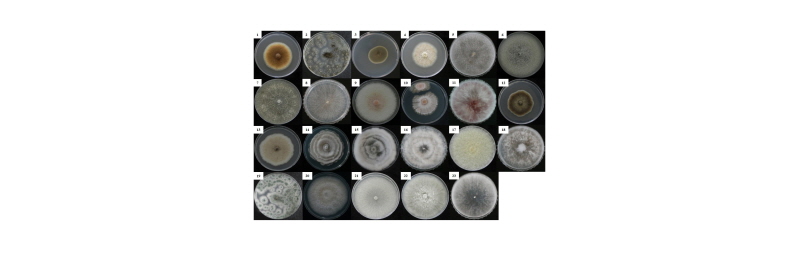
Fig. 2. Colony morphology of the fungal species isolated in this study. Cultures were grown on potato dextrose agar at 25℃ for 7 days. 1, Alternaria alternata; 2, Aspergillus fumigatus; 3,Cladosporim oxysporum; 4, Coprinellus radians; 5, Curvularia buchloes; 6, Daldinia childiae; 7, Daldinia pyrenaica; 8, Discosia pseudoartocreas; 9, Epicoccum nigrum; 10, Eupenicillium saturniforme; 11, Gibberella zeae; 12, Leptosphaeria spegazzinii; 13, Magnaporthe oryzae; 14, Mortierella sclerotiella; 15, Mortierella sossauensis; 16, Mortierella verticillata; 17, Mucor racemosus; 18, Nigrospora oryzae; 19, Penicillium verruculosum; 20, Stagonosporopsis cucurbitacearum; 21, Trichoderma atroviride; 22, STrichoderma hamatum; 23, Trichoderma hispanicum.
한편 황진리에서 채집한 식물체 시료인 사시나무에서 분리한 균주는 버섯 종인 Coprinellus radians로 동정되었으며, 복숭아나무에서 분리된 3개 균주는 버섯 종인 C. radians를 비롯하여 Alternaria alternata, Epicoccum nigrum로 동정되었다. 은사시나무에서 분리된 2개 균주에서도 버섯 종인 C. radians가 Curvularia buchloes와 같이 동정되었다. 흥미롭게도 이들 3가지 서로 다른 식물체에서 버섯 종인 C. radians가 분리 동정된 것은 근처에서 이 버섯의 분포가 많아 포자가 유입되어 식물체 표면에 많이 부착되어 나타난 결과로 보여 진다. 향후 버섯 다양성 조사도 이루어진다면 보다 폭 넓은 진균 다양성이 확인 될 것으로 생각된다. 달뿌리풀에서 분리한 3개 균주는 Alternaria alternata, Nigrospora oryzae, Trichoderma hispanicum로 동정되었다. 물박달나무 시료에서 분리한 3개 균주는 Epicoccum nigrum, Nigrospora oryzae, Mucor racemosus로 동정되었다. 벼과에 속하는 바랭이에서 분리된 균주는 벼도열병을 일으키는 식물병원균인 Magnaporthe oryzae로 동정되었다. 토양시료에서 분리된 균주는 토양의 대표적 균류 중 하나인 Trichoderma atroviride로 동정되었다(Table 1).
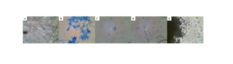
동정된 전체 진균 종 중에 5개 균주(마전리 4균주, 황진리 1균주)는 국내에서 보고가 되어 있지 않은 미기록 진균임을 확인하였다. 이들은 여뀌에서 분리된 Mortierella sclerotiella, 토양에서 분리된 Eupenicillium saturniforme, M. sossauensis, M. verticillate, 달뿌리풀에서 분리된 Trichoderma hispanicum 등이다. 이들 5개 미기록 균주에 대하여 광학현미경으로 관찰한 형태는 Fig. 3과 같다. Mortierella sclerotiella의 균총 형태와 색은 Fig. 2-14과 같다. 25℃, PDA에서 7일 배양한 뒤에 관찰하였을 때 흰색의 균사체가 비슷한 간격으로 층을 내어 가며 자라는 형태를 보였다. 광학현미경으로 균사 끝에 타원형의 포자낭을 관찰할 수 있었으며, 그 크기는 약 1.0~1.5 × 3.5~4.5 μm이었다(Fig. 3A). Eupenicillium saturniforme의 균총은 PDA에서 5일 간 배양하였을 때 연한 핑크색의 균사로 생장하였으며, 균사 말단에 핑크 또는 오렌지색의 균사로 존재하였다(Fig. 2-10). 광학현미경으로 관찰한 결과 3개의 분생자경의 끝에 구형의 포자 (0.5~1.0 × 0.5~0.8 μm)가 체인으로 생장하였다(Fig. 3B). M. sossauensis는 균총은 25℃, PDA에서 7일 배양한 뒤에 관찰하였을 때 아이보리색의 균사체가 비슷한 간격으로 층을 내어 가며 자랐으며 M. sclerotiella와 유사하였다(Fig. 2-15). 광학현미경으로 균사 끝에 타원형의 포자낭을 관찰할 수 있었으며, 그 크기는 약 3.0~3.5 × 5.5~8.5 μm였다(Fig. 3C). M. verticillate는 Fig. 2-16과 같이 25℃, PDA에서 7일 배양하였을 때 M. sclerotiella와 M. sossauensis 처럼 흰색의 균사체가 간격으로 층을 내어가며 자라는 형태를 보였으나 약간 더 부유하여 생장하였다. 광학현미경으로 관찰하였을 때 약 3.0~3.5 × 5.5~6.5 μm의 포자낭을 가지고 있었다(Fig. 3D). Trichoderma hispanicum는 흰색의 균사로 생장하며 말단으로 생장할수록 균사가 부유하는 것을 알 수 있었다(Fig. 2-23). 광학현미경으로 관찰한 결과 3개로 분지한 분생자 끝에 타원형의 분생자경(약 1.0~1.5 × 2.5~3.0 μm)이 존재하였다(Fig. 3E). 이들 생물자원의 활용을 위해 Mortierella sclerotiella는 NIBRFGC000499878, Eupenicillium saturniforme는 NIBRFGC000499890, M. sossauensis는 NIBRFGC000499886, M. verticillate는 NIBRFGC000499888, Trichoderma hispanicum은 NIBRFGC000499887의 번호를 부여받아 국립생물자원관에 생체시료를 기탁하였다.
비무장지대는 서해의 습지와 농경지부터 중·동부의 고산지대를 거쳐 동해안에 이르기까지 한반도 중앙을 가로지르는 248 km 길이의 온대지역이다. 한국전쟁 이후 60여 년 동안 민간인의 접근이 금지 또는 제한된 연유로 비무장지대와 그 주변 일대는 백두대간, 서해안과 함께 한반도의 3대 생태축을 이루는 생태계의 보고로 알려져 있다. 그러나 아직 국내에 보고된 균류에 대한 정보는 거의 없는 실정이다. 본 연구는 집중적인 조사는 아니지만 비무장지대 근역의 하나인 연천군 지역의 산과 강변에서 처음으로 총 18속 23종의 진균을 분리하는 성과를 거두었다. 이는 매우 극히 제한된 식물체와 토양 시료로부터 얻어진 결과이기 때문에 지속적인 조사를 수행한다면 보다 더 다양한 진균에 대한 정보를 얻을 수 있을 것이다. 나아가 향후 기후변화와 생물다양성의 연구를 위해 비무장지대 균류 자원의 탐색에 기여할 수 있을 것으로 생각된다.
적요
비무장지대 일원의 진균 다양성을 연구하기 위한 일환으로 경기도 연천군 마전리에 있는 산과 황진리에 있는 임진강변에서 11가지 식물체 시료와 토양시료 2개를 채집하여 진균을 분리·동정하였다. 그 결과 총 18속 23종의 진균을 확인하였다. 이 중 Eupenicillium saturniforme, Mortierella sclerotiella, M. sossauensis, M. verticillate, Trichoderma hispanicum 등 5종의 진균이 국내에 보고가 되어있지 않는 미기록 종이었다. 본 연구에서 이들의 형태적, 분자적 특성을 기술하였다.
