Novel Fungal Species Belonging to the Genus Acaulium Isolated from Riptortus clavatus (Heteroptera: Alydidae) in Korea

Ju-Heon Lee1   Leonid N.  Ten1   Seung-Yeol  Lee1,2   Hee-Young   Jun1,2,*   

1College of Agriculture and Life Sciences, Kyungpook National University, Daegu 41566, Korea
2Institute of Plant Medicine, Kyungpook National University, Daegu 41566, Republic of Korea

Abstract

A survey of insect-associated fungi in Korea revealed a novel fungal strain isolated from the bean bug Riptortus clavatus (Heteroptera: Alydidae). Culturally and morphologically, the fungal strain designated KNUF-20-INY03, shares features with members of the genus Acaulium. Phylogenetic analyses based on the concatenated nucleotide sequences of the internal transcribed spacer regions (ITS) regions and partial sequences of the translation elongation factor 1-alpha (TEF1-α), and β-tubulin (β-TUB), and large subunit of the nuclear ribosomal RNA (LSU) genes showed that the isolate is part of a clade that includes other Acaulium species, but it occupies a distinct phylogenetic position. Based on the shape, size, and color of its conidia and conidiogenous cells, strain KNUF-20-INY03 is readily distinguishable from the closely related A. acremonium, A. albonigrescens, A. caviariformis, A. pannemaniae, and A. retardatum. The conidial length-to-width ratio (1.6) of the novel isolate is significantly lower than that of A. acremonium (1.9), A. albonigrescens (2.4), and A. pannemaniae (2.4), and KNUF-20-INY03 produces hyaline conidia and elliptical conidiogenous cells while A. caviariformis forms brown conidia and A. retardatum produces flask-shaped conidiogenous cells. Thus, both phylogenetic and morphological analyses indicate that this strain is a novel species in the genus Acaulium, and we propose the name Acaulium microspora sp. nov.

Figures & Tables

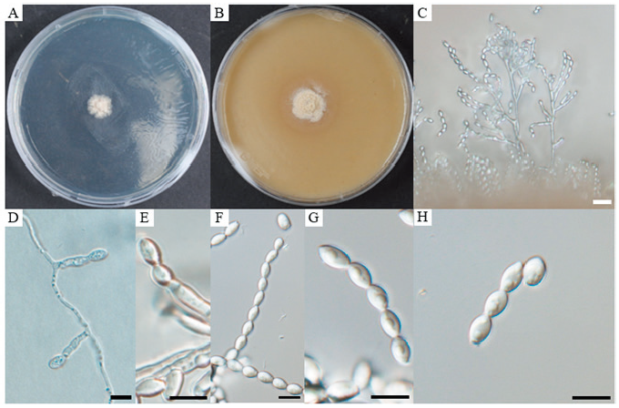
Fig. 1. Cultural and morphological characteristics of . A, colony on potato dextrose agar (PDA) after 14 days of growth at 25℃; B, colony on oatmeal agar (OA) after 14 days of growth at 25℃; C, D, and E, conidiophores and conidiogenous cells; F, G, chain of conidia; H, conidia. Scale bars: C = 20 μm; D–H = 10 μm.