Lactuca serriola L. (syn. L. scariola L.), commonly known as prickly lettuce, is an annual plant belonging to the family Asteraceae. This plant is native to Europe and has become widely naturalized in temperate regions worldwide. It poses a severe risk to agricultural systems, especially in reduced- or no-tillage cultivation methods, which are commonly used for cereals, pulses, soybeans, and wheat [1,2]. L. serriola causes a drastic decline in crop yield by 60-80% at high population density and significantly affects the quality of grains during harvest [1,3,4].
Prickly lettuce was accidentally introduced to Korea in the late 1970s [5,6] and has since rapidly spread to various agricultural and natural habits across the country [7]. Owing to its adverse effects, the Korean Ministry of Environment has designated this weed as a harmful plant that disturbs the balance of the ecosystem [6]. Prickly lettuce seeds are highly adaptable and able to germinate at temperatures as low as 10℃ albeit at a slower speed [3], contributing to their rapid spread across Korea. Furthermore, the survival of this weed in harsh and barren environments, such as the roadsides, in Korea is facilitated by endophytic bacteria, which increase its drought resistance capacity [8].
In June 2020, we observed leaf blight symptoms on prickly lettuce populations invading several farms in Boryeong-si (36°22'46" N, 126°35'37" E) in Korea, with a disease incidence of 30–40% among the inspected plants. Disease symptoms began as small light yellow-to-brown adaxial leaf spots, which gradually coalesced into larger irregular lesions and eventually withered (Fig. 1A and B). Twenty symptomatic leaves were collected and used for fungal isolation. Sections (5×5 mm) of these leaves were surface-sterilized (70% ethanol for 1 min, 0.05% NaOCl for 30 s, and 70% ethanol for 1 min), rinsed twice with sterile water, and placed on a potato dextrose agar (PDA) plate with streptomycin (50 µg/ mL). The plant sections were incubated in the dark at 25℃ until fungal growth was observed. After three days, 15 morphologically similar isolates were selected, and a pure culture was obtained by transferring single hyphal tips to a new PDA plate. Colonies on PDA appeared gray-to-dark brown and velvety, with approximately 4 cm in diameter after three days (Fig. 1C). A sample culture was deposited in the Korean Agricultural Culture Collection (KACC 49814).
The causative agent was identified through a combination of morphological and molecular sequence analyses. Detailed microscopic investigations were performed on the conidiophores and conidia formed on KACC 49814 PDA. Samples were examined and imaged using the Zeiss Imager M2 AX10 microscope (Carl Zeiss, Jena, Germany) equipped with an AxioCam 512 camera (Carl Zeiss). Conidiophores were brown-to-pale brown, simple but rarely branched, septate, thick-walled, and measured (53–) 80 to 128 (–170)×(3–) 4 to 7 (–8) µm (n=30). Conidia (Fig. 1D-F) were dark brown, obclavate to ellipsoidal or fusiform, mostly 3-septate, straight or slightly curved, with a rough verrucose surface, and measured (23–) 28 to 33.3 (–37)×(10–) 12 to 15 (–17) µm (n=50). Terminal cells were smaller, paler, and less verrucose than the central cells. Their morphological characteristics matched those of Curvularia intermedia Boedijn [9].
To confirm the morphological identification, molecular sequence analysis was performed. Genomic DNA was extracted from the fungal conidia and hyphae harvested from KACC 49814 using the DNeasy Plant Mini Kit (Qiagen, Germantown, MD, USA). Then, the internal transcribed spacer (ITS) and glyceraldehyde-3-phosphate dehydrogenase (gpd) [10] were PCR-amplified and sequenced using a DNA sequencing service (Macrogen, Seoul, Korea) with the primers used for amplification. The resulting sequences were edited using the DNASTAR Lasergene software package (DNASTAR, Madison, WI, USA) and deposited in GenBank (accession nos. MZ373300 for ITS and MZ435744 for gpd). A BLASTn search revealed that the Korean isolate had high sequence similarities of 100% and 99.6% with C. intermedia CBS 334.64 for ITS (HG778991) and gpd (LT715828), respectively.
To confirm the morphological identification, molecular sequence analysis was performed. Genomic DNA was extracted from the fungal conidia and hyphae harvested from KACC 49814 using the DNeasy Plant Mini Kit (Qiagen, Germantown, MD, USA). Then, the internal transcribed spacer (ITS) and glyceraldehyde-3-phosphate dehydrogenase (gpd) [10] were PCR-amplified and sequenced using a DNA sequencing service (Macrogen, Seoul, Korea) with the primers used for amplification. The resulting sequences were edited using the DNASTAR Lasergene software package (DNASTAR, Madison, WI, USA) and deposited in GenBank (accession nos. MZ373300 for ITS and MZ435744 for gpd). A BLASTn search revealed that the Korean isolate had high sequence similarities of 100% and 99.6% with C. intermedia CBS 334.64 for ITS (HG778991) and gpd (LT715828), respectively.
Pathogenicity assays were performed twice by spraying a conidial suspension (1.9×10⁵ conidia/mL) onto each leaf of 20 healthy Lactuca serriola (four-week-old) plants. Twenty control plants were sprayed with sterile water. All plants were kept in a growth chamber at 23℃ and 80% relative humidity under a 12-h day/night cycle. One week after inoculation, initial symptoms began to emerge as small foliar spots, which turned yellow to brown in two weeks, leading to leaf dieback and necrosis (Fig. 1G-I). Control plants
Fig. 1
Leaf blight on prickly lettuce (Lactuca serriola) caused by Curvularia intermedia. (A and B) Symptoms observed on the upper (A) and lower (B) surfaces of an infected leaf. (C) A close-up of a leaf blight lesion on the upper leaf surface. (D) Colony morphology observed on the potato dextrose agar (PDA) plate after incubation for a week in darkness at 25℃. (E-G) Conidia. (H and I) Pathogenicity assays showing typical brown spots (arrows) on the front (H) and backside (I) of an inoculated leaf. (J) Enlarged region of the inoculated leaf showing symptoms. (K) Conidia re-isolated from an inoculated leaf. Scale bars: 10 µm (E-G) and 30 µm (K).
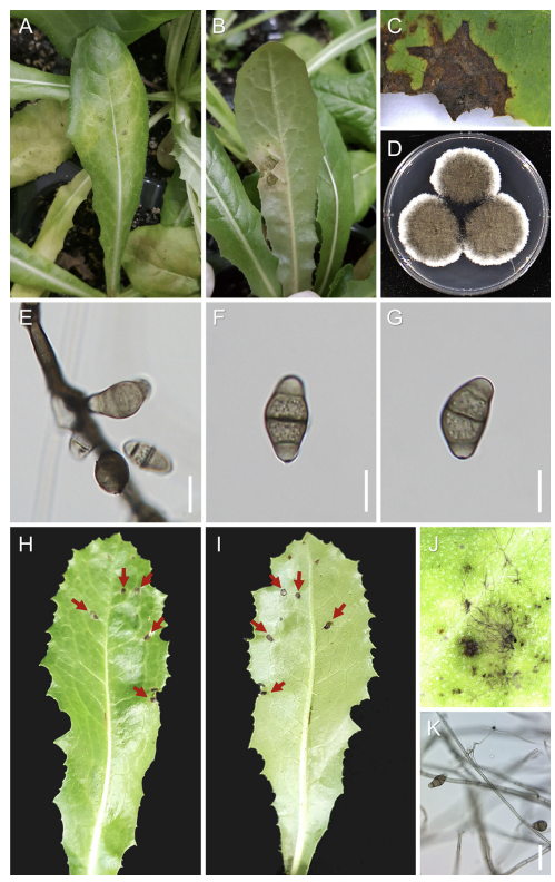
were symptom-free. The same fungus was successfully reisolated from the artificially inoculated plants (Fig. 1J), thereby fulfilling Koch’s postulates.
In the last decade, several fungal pathogens, including Alternaria alternata [11], Golovinomyces orontii (recently reclassified under G. bolayi) [12], Fusarium fujikuroi [13], and Septoria lactucae [14], have been reported on L. serriola in Korea. Additionally, oomycetes, such as Bremia lactucae [15], have also been documented. To the best of our knowledge, this is the first report of leaf blight caused by C. intermedia on L. serriola in Korea. C. intermedia affects the economically important crops and grasses as well as weeds of lawns and turfgrasses [16]. Furthermore, it causes leaf spots on Zea mays in Korea [17].
Results of this study suggest C. intermedia as an effective biocontrol agent for L. serriola. This pathogen was previously proposed as a potential microbial herbicide for large crabgrass (Digitaria sanguinalis) [18]. In addition, use of C. intermedia as a microbial herbicide against crabgrass and other susceptible species has been detailed in two U.S. patents [19,20]. However, its potential non-target effects require further investigation to prevent any adverse ecological impact.

