Abstract
Figures & Tables
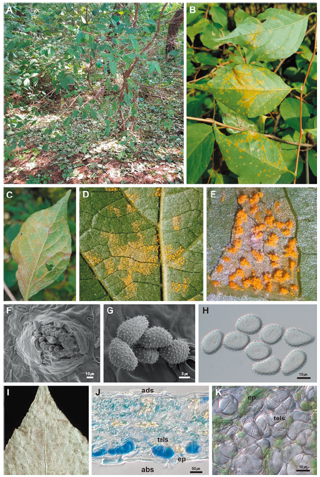
Fig. 1. occurring on . A, early defoliation of an affected tree in late September, 2020. B, symptoms on the lower leaf surface of affected leaves in the early stage. C, symptoms on the lower leaf surface in the advanced stage. D and E, uredinia formed on the lower leaf surface. F and G. Uredium (F) and urediniospores (G) observed under a scanning electron microscope. H, urediniospores focused on outline under a DIC microscope. I, symptoms on the lower leaf surface with the telial stage. J, vertical section of teliospores on the leaf epidermis on the abaxial side. K, cross section of teliospores seen from the leaf epidermis. abs, abaxial side; ep, epidermis of the leaf; tells, teliospores.